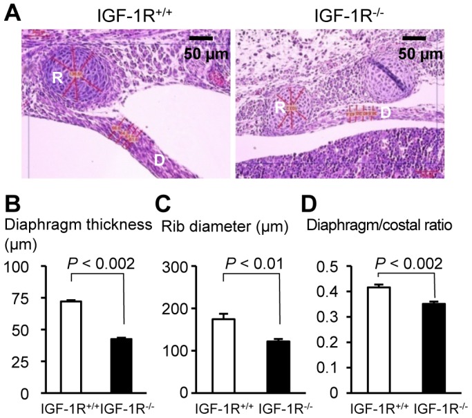
Figure 6. Development of diaphragm and chest in the absence of IGF-1R.

A, Hematoxylin-eosin stained transversal section of thoracic wall and diaphragm in control (left) and IGF-1R−/− embryos (right) at E17.5. Bar graphs compare B, diaphragm thickness, C, rib diameter, and D, diaphragm-to-rib ratio (mean ± SEM) in IGF-1R+/+ embryos (n = 4) and IGF-1R−/− embryos (n = 4). R, Rib; D, diaphragm. Wilcoxon Mann-Whitney U test.
