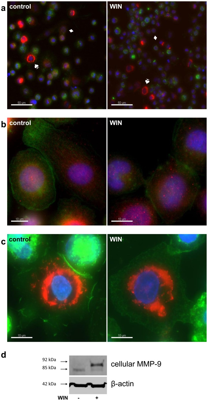
Figure 4. WIN did not alter the intracellular localization of MMP-9.
Immunocytochemical staining of MMP-9 (red), F-actin (green) and nuclei (blue) of U937-macrophages which were treated for 24 h with 4 µM WIN or DMSO (control). The figure shows one representative analysis out of five. (a) Two types of MMP-9 expressing cells are observed in control cells and in WIN-treated cells, cells with a low MMP-9-signal in a vesicular pattern (♦), and cell with a brighter signal near the nucleus (♠). (b) Magnification of the cell type with MMP-9 in vesicular distribution. (c) Magnification of the cells type with perinuclear MMP-9-distribution. (d) Western blot analysis of the same samples using the same MMP-9 antibody as for immunocytochemistry (Abcam ab38904).

