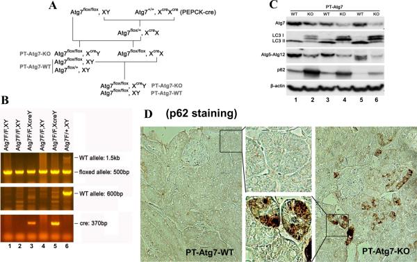
Figure 3. Creation and characterization of the PT-Atg7-KO) mouse model.
(A) Breeding protocol for generating PT-Atg7-KO mice. Male littermate mice of 8–9 weeks old were used for experiments after genotypes confirmed. (B) Representative images of PCR-based genotyping. Genomic DNA was extracted from tail biopsy and amplified to detect wild-type (WT) and floxed alleles of Atg7 and PEPCK-Cre allele as indicated. (C) Whole tissue lysate of kidney cortex was collected from PT-Atg7-KO and wild-type (PT-Atg7-WT) littermate mice for immunoblot analysis of Atg7, LC3, Atg5 (Atg12 conjugated), p62, and β-actin. (D) Immunohistochemical staining of p62 (×200) in kidney cortical tissues of wild-type and PT-Atg7-KO mice. The selected areas were shown at high magnification in the middle panels.

