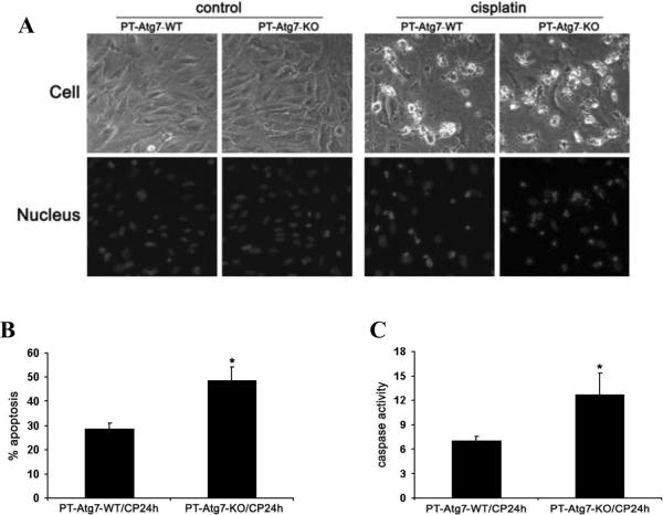
Figure 7. Proximal tubular cells isolated from PT-Atg7-KO mice are sensitized to cisplatin-induced apoptosis.
Primary proximal tubular cells isolated from wild-type and PT-Atg7-KO mice were treated with 30μM cisplatin for 24 hours. Apoptosis was assessed by cell morphology and caspase activation. (A) Representative cell and nuclear morphology (×200). After treatment, cells were stained with Hoechst33342 to record cell and nuclear morphology. (B) Apoptosis percentage. Apoptotic cells were counted to determine the percentage of apoptosis. (C) Caspase activity measured by enzymatic assays using carbobenzoxy-Asp-Glu-Val-Asp-7-amino-4-trifluoromethyl coumarin as substrates. Data in (B) and (C) are expressed as mean ± SD. * P < 0.05, significantly different from the wild-type group.

