
Fig. 1.
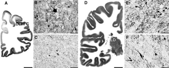
GAD 65/67 immunoreactivity in the left DLPFC (dorsolateral prefrontal cortex) and the right LD (lateral dorsal thalamic nucleus) of unipolar (b and e, respectively) and bipolar I (c and f, respectively) depressed patients; scale bars 50 μm. These regions of interest are shown at low magnification in pictures of GAD-immunostaining of a control case (a and d; scale bars 10 mm)