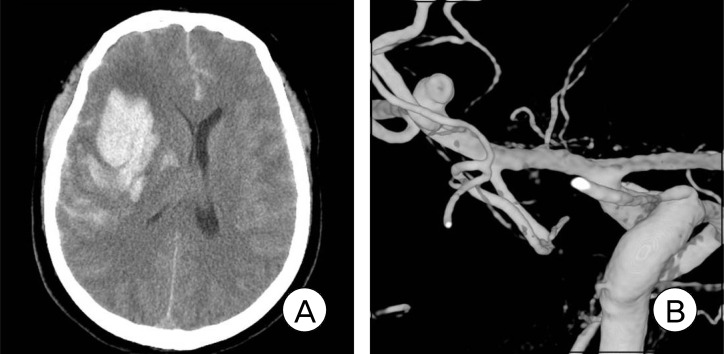
Fig. 1.
Admission brain computed tomography (CT) scan shows right frontal intracerebral hemorrhage (ICH) with a mass effect by ipsilateral ventricle compression (A). Right three-dimensional digital subtraction angiogram (3D-DSA) shows the rupture point of the middle cerebral artery (MCA) bifurcation aneurysm projecting superiorly (B).

