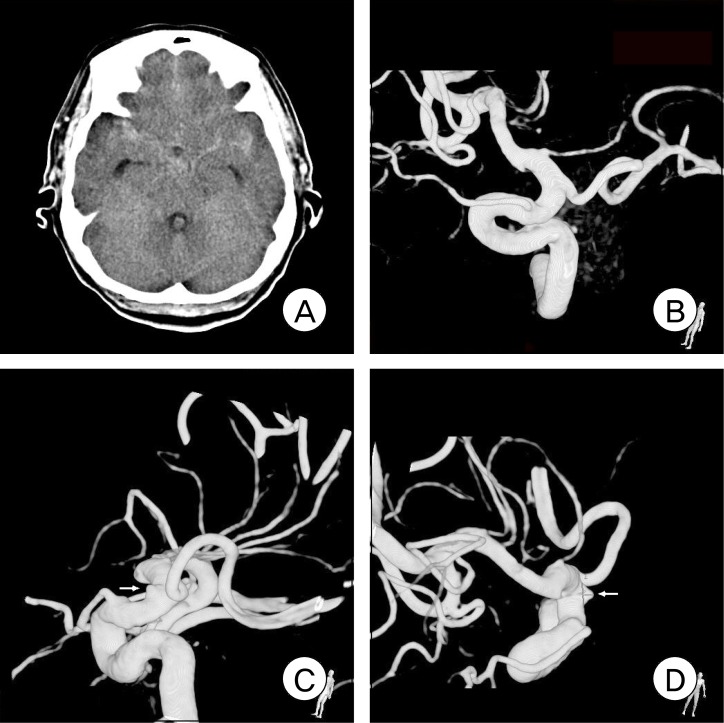
Fig. 1.
Initial noncontrast brain computed tomography (CT) and digital subtraction angiography (DSA). Diffuse subarachnoid hemorrhage (SAH) in the region of both the sylvian fissure and interhemispheric fissure (A). Initial three-dimensional rotational angiography image reveals no aneurysmal structure (B). Repeat DSA was performed on the eighth day of admission. Three-dimensional rotational angiography images clearly show a distal ICA aneurysm (C, D).

