Figure 2.
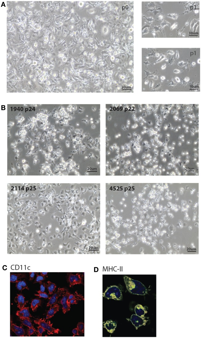
Morphology of MutuDC line cultures. (A,B). Light microscopy images of five representative MutuDC lines at early [(A), MutuDC line 4417] or later passages [(B), MutuDC lines as indicated]. MutuDC lines may show different morphologies (in (B)], e.g., MutuDC1940 tends to show more aggregates and MutuDC2069 presents less dendrites. Moreover, these traits tend to be more pronounced in all cell lines with longer times of culture without splitting or harvest. (C,D) Immunofluorescence microscopy of one representative DC line (MutuDC2114) showing CD11c [in red, in (C)] and MHC-II [in yellow, in (D)]. Nuclei were stained with DAPI.

