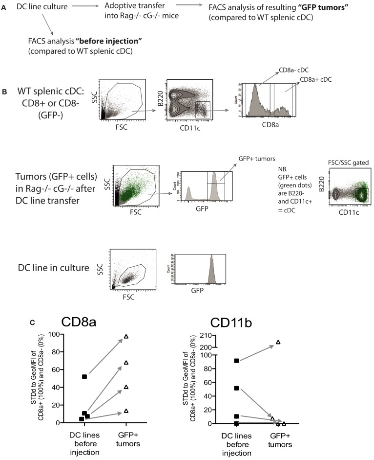
Figure A2.
Most MutuDC lines restore CD8α expression and downregulate CD11b upon adoptive transfer in vivo. (A) Experimental outline: MutuDC lines (n = 4) were analyzed directly from culture or following intra-veinous adoptive transfer of 3 × 10⁶ cells per MutuDC line into immunodeficient rag2−/−γc−/− hosts. Spleens containing MutuDC line cells (tumors) were analyzed 16 days after injection. (B) Gating strategy for the flow cytometry analysis of MutuDC lines in culture versus tumors in MutuDC line-injected mice. MutuDC lines and normal WT splenic cDC subsets (used as controls) were gated as indicated (and as previously described in Figure 4A). MutuDC line cells in tumoral splenocytes from MutuDC line-injected mice were gated based on eGFP expression as indicated. (C) The expression of surface markers CD8α and CD11b was analyzed in MutuDC lines before injection versus following adoptive transfer in vivo and standardized relative to the levels found in normal WT splenic cDC subsets (controls). For CD8α expression, the CD8α+ (CD11b−) cDC subset represents 100% and the CD8α− (CD11b+) cDC subset represents 0%, and vice versa for CD11b expression. Arrows indicate the change in surface marker expression for each particular MutuDC line.

