Figure 2.

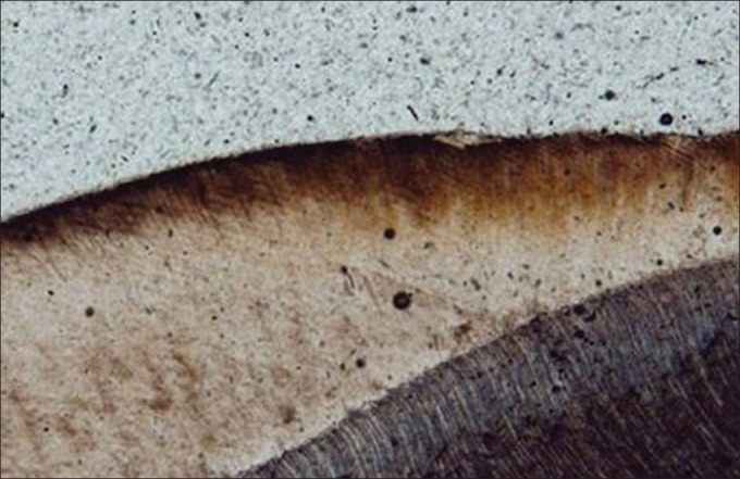
Photomicrograph of enamel lesion representing the acid-etched group at ×40 magnification under polarized light microscope

Photomicrograph of enamel lesion representing the acid-etched group at ×40 magnification under polarized light microscope