
Figure 3.
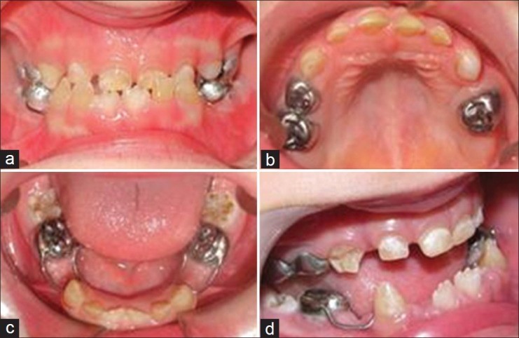
Intraoral view. (a) Frontal view of the occlusion. (b) Occlusal view of the upper arch. (c) Occlusal view of the lower arch after placement of space maintainers. (d) Enamel disintegration on the right upper canine (a lateral view)
Official websites use .gov
A
.gov website belongs to an official
government organization in the United States.
Secure .gov websites use HTTPS
A lock (
) or https:// means you've safely
connected to the .gov website. Share sensitive
information only on official, secure websites.
Intraoral view. (a) Frontal view of the occlusion. (b) Occlusal view of the upper arch. (c) Occlusal view of the lower arch after placement of space maintainers. (d) Enamel disintegration on the right upper canine (a lateral view)