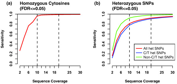
Figure 4.
Sensitivity as a function of sequence coverage. Comparisons between Bis-SNP SNP calls and 1 M SNP array from Figure 3 ROC curves were extended to a range of coverage levels from 2×-30×. At each coverage level, we selected the least stringent threshold that yielded a False Discovery Rate (FDR) less than 0.05, and plotted the Sensitivity (1 - False Negative rate). As in Figure 3, separate plots show sensitivity at detecting homozygous cytosines (a) and heterozygous SNPs (b). For heterozygous SNPs, we include the overall detection rate (red line), as well as separate lines for C/T heterozygous SNPs (blue line) and non-C/T heterozygous SNPs (green line).

