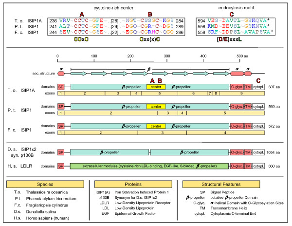
Figure 6.
The low-iron inducible receptor ISIP1. ISIP1 protein models and secondary structure from T. oceanica, P. tricornutum and F. cylindrus are compared. Conservation between the protein orthologs is high, with identical secondary structure predictions (center). We find an amino-terminal signal peptide targeting the protein to the secretory pathway, while a carboxy-terminal transmembrane domain anchors the protein to a membrane. The major part of the protein is represented by a domain rich in β-strands that likely folds into a β-propeller-like structure. While in D. salina p130B (bottom) this β-propeller domain is duplicated and only distantly related to the respective diatom domains, the remainder of the protein shows a clear homology to the group of diatom ISIP1 proteins. A clue to the structure and function of ISIP1 could be the human low-density lipoprotein receptor LDLR due to its detailed characterization as a human cell-surface receptor: while its extracellular domains are very different from the single β-propeller domain of ISIP1, the remainder of the protein is again strikingly similar, which allows us to transfer the respective annotation from LDLR to the ISIP1 protein model. Accordingly, the ISIP1 protein would represent a cell-surface receptor that is anchored to the plasma membrane by a carboxy-terminal transmembrane helix. A small carboxy-terminal tail without well-defined secondary structure contains a conserved endocytosis motif C (top, right) responsible for endocytotic cycling of ISIP1. An α-helical region amino-terminal from the transmembrane helix is predicted to be O-glycosylated and thereby would serve to expose the large β-propeller as a putative receptor domain to the extracellular space. A sequence alignment of the ISIP1 proteins from T. oceanica, P. tricornutum and F. cylindrus illustrates that the extracellular β-propeller domain contains a cysteine-rich center, A and B (top, left). The pattern of cysteine residues is reminiscent of patterns found in Fe-S cluster proteins and might also be involved in binding Fe.

