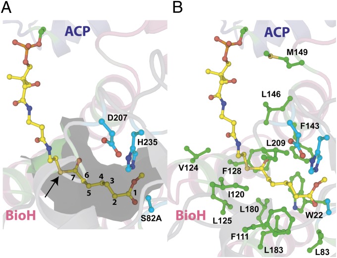
Fig. 4.
A hydrophobic channel directs the methyl pimeloyl moiety toward the active site for catalysis. The structural elements are colored as in Fig. 2. The phosphopantetheine-linked pimeloyl methyl ester is shown in stick-ball representation with carbon atoms colored yellow. (A) Internal cavity around the methyl-pimeloyl thioester at a distance of 4.5 Å is shown as a partially transparent surface colored green. The pantetheine sulfur atom is marked by an arrow, and the seven-carbon pimeloyl chain is traced by numerals against carbon atoms. (B) Side chains of all residues at a maximum distance of 4.5 Å from the phosphopantetheine-linked pimeloyl methyl ester are shown with their side-chain atoms in stick-ball representation with the carbon atoms colored green.

