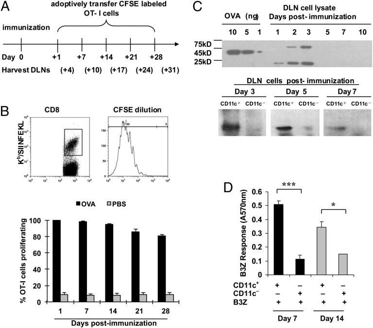
Fig. 1.
OVA persists in vivo for several weeks after immunization. (A) Experimental design for B (Upper). Gating strategy used for CFSE-diluting CD8-positive, Kb/SIINFEKL pentamer-positive cells in drainage lymph nodes (DLNs) of immunized mice in the results shown in the lower panel. (B, Lower) Proliferation of OT-I cells measured by CFSE dilution (shown) in the DLNs of C57BL/6 mice immunized with soluble 200 μg OVA protein (black bars) or phosphate-buffered saline (gray bars). (C) OVA protein or its fragments were detected in the DLN cells (Upper) by sodium dodecyl sulfate polyacrylamide gel electrophoresis followed by immunoblotting with a polyclonal antibody at indicated time points after OVA immunization. Titrated quantities of soluble OVA (10, 5, or 1 ng) were used as quantitative standards in the left lanes. A similar analysis was conducted on CD11c-positive and CD11c-negative cells (72,000 cells each) derived from the DLNs of immunized mice 3, 5, and 7 d after immunization. (D) Stimulation of B3Z cells by CD11c-positive cells purified from DLNs of C57BL/6 mice immunized with soluble 200 μg OVA protein 7 d (black bars) or 14 d (gray bars) after immunization. *P < 0.05; **P < 0.01; ***P < 0.001. Error bars indicate mean ± SD. Experiments were conducted three to five times each. Three to five mice per group were used.

