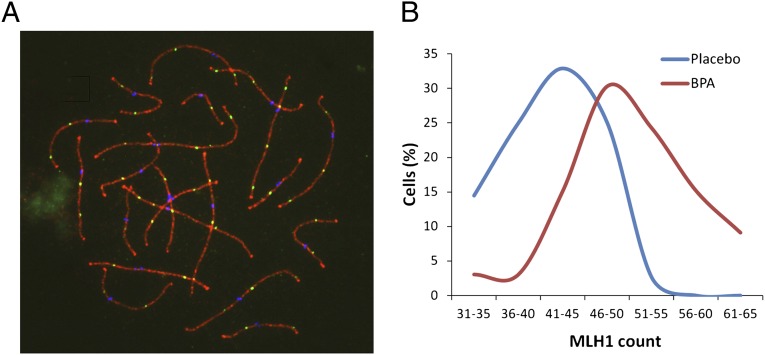
Fig. 1.
Recombination is increased in BPA-exposed females. (A) Example of pachytene oocyte used to obtain MLH1 counts. Triple immunostaining with antibodies to SYCP3 (red), MLH1 (green), and CREST (blue) allowed detection of synaptonemal complex, sites of recombination, and centromeres, respectively. (B) Comparison of MLH1 counts from 33 cells from fetuses continuously exposed to BPA (red curve) and 70 from placebo-treated controls (blue curve). Mean values were highly significantly different (50.4 ± 7.0 and 42.2 ± 5.9 for BPA-exposed and placebo, respectively; t = 6.8; P < 0.001).

