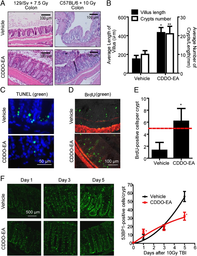
Fig. 6.
CDDO-EA protects the GI tract from acute TBI. (A) Representative images of H&E staining of the colon tissue in different strains at 5 d after TBI with or without prior feeding of CDDO-EA chow. (B) Length of villi (left y axis) were measured in at least 40 complete, well-oriented villi cross-sections in the small intestine of 129/Sv mice 5 d after a 7.5-Gy dose of TBI. Crypt number per 1-mm length of the small intestine was counted (right y axis) (n = 3). (C–F) WT C57BL/6 mice were exposed to 10-Gy TBI 3 d after feeding of control or CDDO-EA chow. Representative TUNEL staining (green fluorescence) in the colon (C; 1 d postirradiation) and immunohistochemical detection of in vivo BrdU incorporation (green fluorescence) in the colon crypts (D; 3 d postirradiation) are shown. DAPI (blue) or smooth muscle actin (red fluorescence) was used for counterstaining. (E) BrdU-positive cells were counted in 15 complete, well-oriented crypt cross-sections (n = 3). The dashed line indicates the number of BrdU-positive cells considered critical for crypt survival (47). (F) Colon tissues were immunostained using anti-53BP1 antibodies 1, 3, or 5 d after a 10-Gy dose of TBI. (Left) Representative images show 53BP1-positive cells in colon tissues. DAPI was used for counterstaining (Fig. S7). (Right) Number of 53BP1-positive cells was counted in at least 60 complete, well-oriented crypt cross-sections in the colon (n = 3). *P < 0.0001 and **P = 0.0016 (compared with vehicle control) in the unpaired Student t test.

