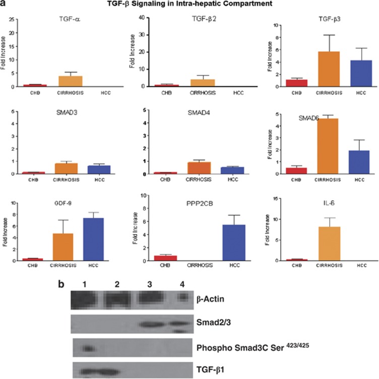
Figure 4.
ABI custom array of 48 genes of TGF-β signaling pathway was designed and quantitative real-time PCR analysis was performed with Syber Green. In peripheral blood mononuclear cells (PBMCs) of chronic HBV (CHB), cirrhosis and hepatocellular carcinoma (HCC) patients in (a) liver infiltrated lymphocytes (LiLs) of CHB, cirrhosis, and HCC patients. Signaling module depicting activated gene sets of TGF-α, TGF-β2, GDF9, SMAD1, SMAD4, MAPK14, BMP6, BMPR2, and PPP2CB in PBMCs from HCC patients, which were attenuated during the cirrhosis stage of infection. (a) In LiLs, increased expression of TGF-α, TGF-β2, TGF-β3 GDF9, SMAD3, SMAD4, SMAD6, and interleukin-6 was observed. (b) Western blot showing β-Actin, TGF-β, Smad2/3, and pSmad3C Ser.423/425

