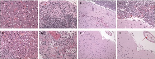
Fig 6.
Anthrax-related lesions in the livers, lymph nodes, and brains of treated and untreated NHPs. (A) Image at ×40 magnification of liver section from a monoclonal antibody-treated NHP that died 3 days after challenge. Mild sinusoidal leukocytosis and a few circulating B. anthracis organisms (arrow) are evident. (B) Image at ×40 magnification of a liver section from an untreated NHP that died 7 days after challenge. A focus of hepatocellular necrosis, fibrin exudation, suppurative inflammation, and numerous intralesional B. anthracis organisms can be seen in the lower left portion of the image. (C) Image at ×40 magnification of mediastinal lymph node from monoclonal antibody-treated NHP that died 3 days after challenge. Mild lymphoid depletion, subcapsular edema, minimal hemorrhage, and a few B. anthracis organisms (arrow) are apparent. (D) Image at ×40 magnification of mediastinal lymph node from an untreated NHP that died 5 days after challenge. Marked lymphoid necrosis, fibrin exudation, hemorrhage, and intralesional B. anthracis organisms (arrow) can be seen. (E) Image at ×20 magnification of the cerebral cortex from a monoclonal antibody-treated NHP that died 4 days after challenge. Suppurative meningitis, vasculitis, hemorrhage, and necrosis (n) are visible. (F) Image at ×20 magnification of the meninges and cerebral cortex from an untreated NHP that died 3 days after challenge. Note the typical lack of notable microscopic lesions. (G) Image at ×40 magnification of the cerebral meninges from a monoclonal antibody-treated NHP that died 4 days after challenge. Suppurative meningitis and hemorrhage with intralesional B. anthracis organisms (arrow) are evident. (H) Image at ×40 magnification of the cerebral meninges from an untreated NHP that died 3 days after challenge. Note the presence of intravascular B. anthracis organisms (arrow) but no other microscopic lesions. hematoxylin and eosin stain was used for all tissues.

