Figure 3.
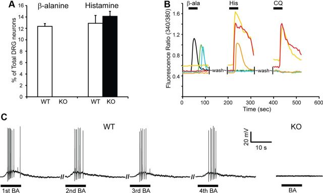
The response of DRG neurons to β-alanine is MrgprD-dependent. A, Approximately 12% of WT DRG neurons responded to β-alanine (β-ala; 1 mm) with increased [Ca2+]i, whereas MrgprD−/− DRG neurons did not respond (number of neurons analyzed: WT, n = 282; KO, n = 212, n = 3 mice per genotype). The response to histamine was not impaired in MrgprD−/− neurons. The percentage of MrgprD−/− neurons responding to histamine (100 μm) was similar to that of WT neurons (>300 neurons analyzed, 3–4 mice per genotype). Error bars represent SEM. B, Representative traces of DRG neurons from MrgprDGFP/+ mice in calcium imaging assays. β-alanine only activated GFP+ neurons as monitored by increased [Ca2+]i with calcium imaging. β-alanine-sensitive neurons (black, green, and blue traces; n = 91) did not respond to histamine (His; 100 μm) or CQ (1 mm), and histamine- and CQ-sensitive neurons (yellow, red, and orange traces) did not respond to β-alanine (>200 neurons from 3 mice). C, β-alanine (BA; 1 mm) induced action potentials in DRG neurons. In WT DRG, β-alanine-sensitive neurons (as determined by calcium imaging, n = 8) fired action potentials upon repeated β-alanine treatment. In contrast, none of the neurons tested from MrgprD−/− mice showed any response to the drug (n = 7).

