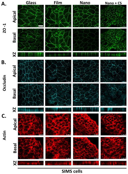
Figure 5.
PLGA nanofibers promote apical restriction of ZO-1, which is disrupted by chitosan. Immunostaining detected (A) ZO-1 and (B) occludin localization. (C) The actin cytoskeleton was detected using rhodamine-phalloidin. Representative single confocal (63×) images were selected from Z-stacks to represent apical and basal sections of SIMS cells on the basis of β-actin and integrin α6 localization (data not shown). XZ projections provide a vertical view of protein localization throughout the height of the cell. SIMS cells seeded on nanofibers fibers showed restriction of ZO-1 but not occludin to the apical side of the cell, which is indicative of AJ but not mature TJ formation. Chitosan-modified nanofiber scaffolds disrupted the apical restriction of ZO-1. Scale, 25 μm.

