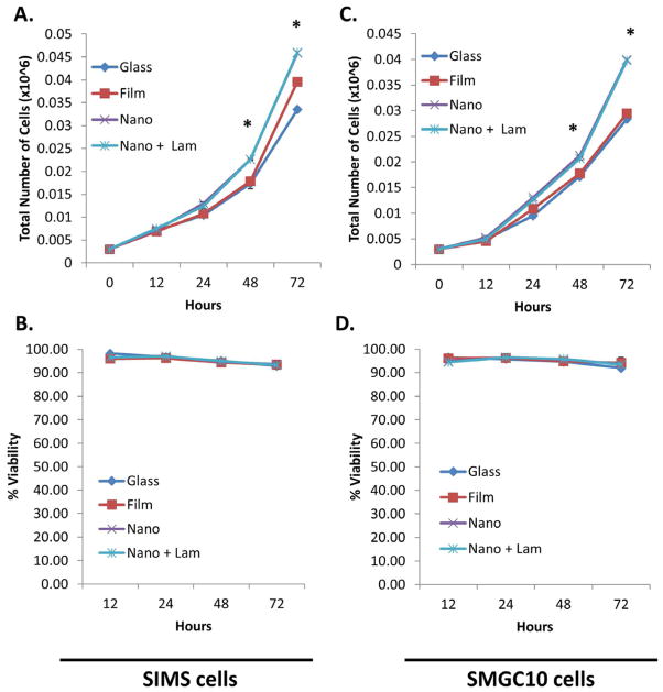
Figure 8.
Laminin-111-modified nanofibers increase ductal (SIMS) and acinar (SMGC1O) cell proliferation without affecting cell viability. Total cell number graphs showing increased (A) SIMS cell and (B) SMGC10 cell proliferation for cells cultured on laminin-111-modified scaffolds as compared to unmodified nanofibers and flat substrate controls from 12–72 hrs. Mean ± SEM of 2 experiments. One-way ANOVA with Bonferroni post-tests indicates a significant difference (* p<0.05) between nanofibers (unmodified and laminin-111-modified) and flat substrates (glass and PLGA film) at 48 and 72 hours. Cells cultured on laminin-111-modified vs. unmodified nanofibers did not show a difference in proliferation rate. Graphs for % viability of (C) SIMS and (D) SMGC10 cells show that viability is not affected by the laminin-111 coating. Mean ± SEM of 2 experiments. One-way ANOVA with Bonferroni post test results indicate no significant difference in viability (p>0.05) for all substrate types at all time points.

