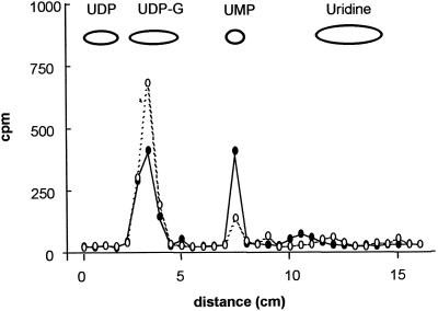
Figure 4.
Metabolism of [3H]UDP-Glc by pea stem Golgi vesicles. Golgi vesicles (100 μg of protein) were incubated with [3H]UDP-Glc (3 μCi/nmol) for 10 min (•). As a control, Golgi vesicles previously boiled for 3 min were incubated under the same conditions (○). After 10 min an aliquot of each sample equivalent to 2 μg of protein was taken, boiled for 1 min, and loaded onto a PEI-TLC plate. The plate was run, air-dried, and cut into 0.5-cm fragments. The radioactivity associated with each fragment was determined by liquid-scintillation counting. The migration of standards is depicted.

