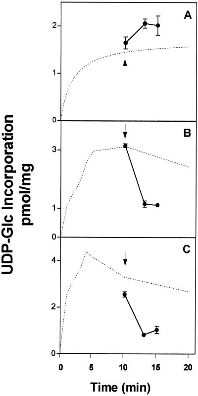
Figure 8.
UDP-Glc-induced efflux of radiolabeled solutes associated with Golgi vesicles. Golgi vesicles (300 μg of protein) were incubated for 10 min with 1 μm (3 μCi/nmol) [β-32P]UDP-Glc (A), [α-32P]UDP-Glc (B), or [3H]UDP-Glc (C). After the incubation a pulse of cold UDP-Glc (1 mm) was added to each sample (indicated with an arrow), and the incubation continued for 0, 3, or 5 min. Finally, the incubations were stopped by filtration, and the radioactivity that remained associated with the vesicles after the pulse of UDP-Glc was determined by liquid-scintillation counting. The amount of solutes associated with the vesicles is expressed in equivalents of UDP-Glc at the beginning of the incubation. The measurements were done in triplicate and the average and its deviation are depicted in the figure. The incorporation of the various substrates, obtained from Figure 6, is depicted with a broken line.

