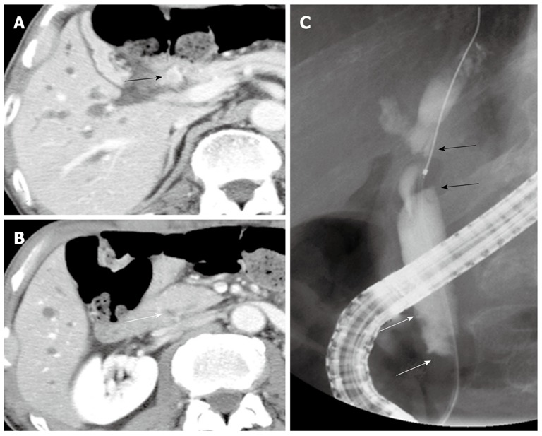
Figure 1.

Imaging findings before the first surgery. A: Computed tomography showed a well enhanced mass in the middle and superior parts of the bile duct (black arrow); B: No mass was detected in the inferior part of the bile duct (white arrow); C: Endoscopic retrograde cholangiopancreatography revealed a tuberous filling defect in the middle and superior parts of the bile duct (black arrows) and mild stenosis in the inferior part (white arrows).
