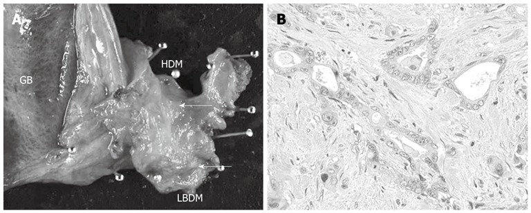
Figure 2.

Macroscopic and microscopic findings of resected specimen at the first surgery. A: Resected specimen of the extra-hepatic bile duct showed whitish tuberous tumor in the middle and inferior parts of the bile duct (cancer marked by the white arrows); B: Microscopically, the tumor was moderately differentiated tubular adenocarcinoma with invasive growth (hematoxylin-eosin; magnification; × 100). GB: Gallbladder; HDM: Hepatic duct margin; LBDM: Lower bile duct margin.
