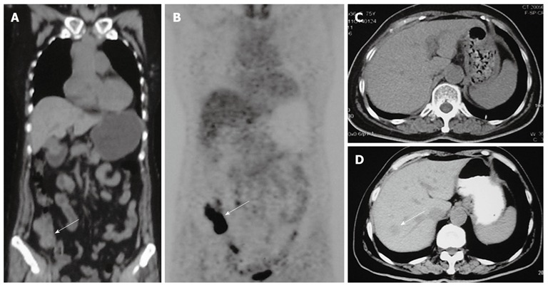
Figure 1.

Positron emission tomography plus computed tomography and computed tomography scan findings in our patient. A: Positron emission tomography plus computed tomography (PET/CT) scan showed thickening of the ileocecal region (white arrow), and no liver lesions; B: PET/CT scan showed increased metabolic areas in the ileocecal region (white arrow), no increased metabolic areas in the liver; C: Six months later a follow-up CT scan showed no liver lesions; D: Eleven months later a follow-up CT scan showed one low-density lesion in the right liver lobe (white arrow).
