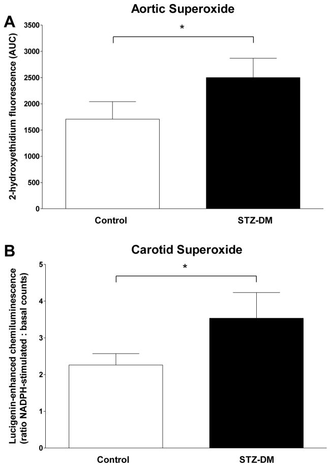
Figure 5. Arterial superoxide levels in control and streptozotocin-treated monkeys.
Segments of carotid and aortic arteries were assessed for superoxide levels as outlined in Methods. Panel A: data are the mean ± SEM for superoxide anion-induced 2-hydroxyethidium fluorescence in aortic segments isolated from control (white bars; n = 9) and STZ-DM monkeys (black bars; n = 9). Arterial rings were incubated in dihydroethidine and subjected to HPLC separation. Formation of 2-hydroxyethidium in the presence of superoxide anion was quantified by fluorescence area-under-the-curve. Panel B: data are the mean ± SEM for the ratio of NADPH-stimulated to basal chemiluminescence in carotid arteries isolated from control (white bars; n = 9) and STZ-DM monkeys (black bars; n = 9). Luminescence is produced by the reaction of lucigenin with superoxide anion in the presence of NADPH. (* = P < 0.05).

