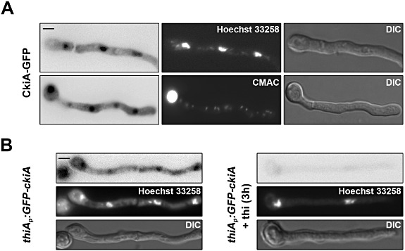
Fig 5.

Intracellular localization of CkiA–GFP. A. Localization of CkiA–GFP in germlings. Two germlings are shown, the top one is counterstained with Hoechst 33258, the bottom one with CMAC. Germlings of strain VIE172 grown in MM for 16 h on urea as sole nitrogen source. B. Epifluorescence detection of GFP-CkiA driven by the thiA promoter in germlings (strain VIE047) grown in MM for 14 h on proline as sole nitrogen source and then shifted to proline in the absence and presence of thiamine (thi) for the additional time indicated. Counterstaining with Hoechst 33258 is also shown. Scale Bars: 5 µm.
