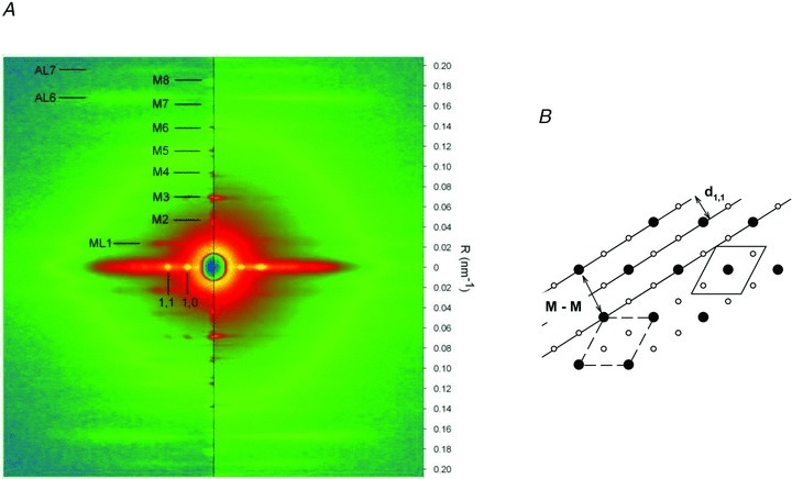
Figure 1. X-ray diffraction.

A, two-dimensional diffraction patterns from dogfish muscle fibres at rest (left half) and at tetanus plateau (right half). The vertical axis (meridional) is parallel to the fibre axis and the scale on the right indicates the position in reciprocal space (nm−1). The myosin-based meridional reflections are indexed as M1 … M8. ML1, AL6 and AL7 indicate the first myosin-based layer line and the actin-based layer lines. The horizontal (equatorial) axis (perpendicular to the fibre axis) contains the (1,0) and (1,1) reflections. The two patterns are the sum of 5 ms frames from 3 fibre bundles, for a total exposure time of 105 ms at rest and 140 ms at the plateau of the isometric tetanus. B, diagram of the hexagonal array of thick and thin filaments in the myofilament lattice. The distance d1,1 between the continuous lines (representing the 1,1 planes) is half the myosin-to-myosin spacing (M-M). The unit cell, containing one thick filament, is shown by the dashed line. The shifted version of it, continuous line, makes more explicit the ratio 1:2 between myosin and actin filaments.
