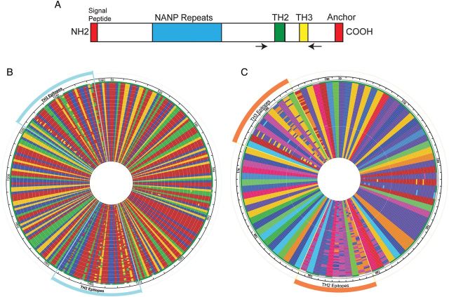
Figure 1.
TH2 and TH3 polymorphisms and haplotypes of csp variants. A, Schematic structure of CS (not to scale) with the locations of the NANP repeat (blue), TH2 epitope (green), and TH3 epitope (yellow) highlighted. The signal peptide and anchor sequences are also shown (red). The locations of the primers are marked by the arrows. B, DNA alignment of all 57 unique variants detected in the population. Nucleotides are represented by different colors (adenine, red; thymine, blue; cytosine, green; and guanine, yellow). The position of the TH2 and TH3 epitopes are marked. C, Amino acid alignment of all 57 unique variants detected in the population. Amino acids are represented by different colors (alanine, brick red; arginine, yellow ochre; asparagine, violet; aspartic acid, orange yellow; cysteine, forest green; glutamic acid, blue; glutamine, medium blue; glycine, dirty yellow; histidine, bright red; isoleucine, red; leucine, dark purple; lysine, pink; methionine, magenta; proline, indigo; serine, light blue; threonine, bright blue; tryptophan, medium green; tyrosine, parrot green; and valine, light green; phenylalanine is not seen). The position of the TH2 and TH3 epitopes are marked.

