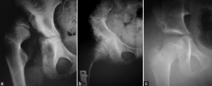
Figure 6.

Anteroposterior radiograph of a 5.8-year-old girl (case 16) showing (a) unilateral dislocation of the hip right side; (b) followup radiograph 7 months after surgery with adequate reduction; (c) followup radiograph after 7.6 years of surgery, with well-developed congruous hip joint
