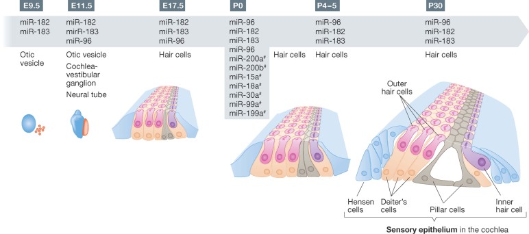
Figure 1. Timeline for miRNA expression during development and early postnatal stages of the inner ear for a subset of miRNAs.
The most well-studied miRNAs, mir-96, -182- and -183, were first detected in the otic vesicle at E9.5, progressed to the cochlea-vestibule ganglion and the neural tube at E11.5, and by E17.5 had strong expression in the cochlear hair cells. The expression continued to at least p30, by some reports. The expression of other miRNAs detected by in situ hybridization are shown as well. Those marked with # were only examined at the stage indicated.

