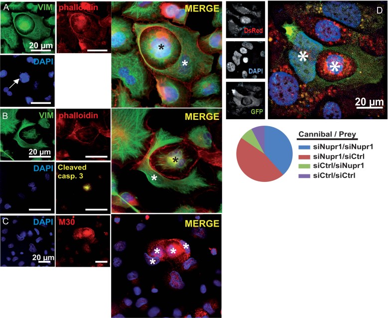
Figure 4. Nupr1-depletion is permissive for TGFβ-stimulated PDAC cell cannibalism and subsequent cell death.
Fluorescence microscopy pictures of Nupr1-depleted cells after 48 h of TGFβ stimulation.
- Actin (red as in A and B) and vimentin (green) immunostaining. Note the nuclear fragmentation of the inner cell (arrow).
- Cleaved-caspase 3 (yellow) staining.
- Caspase-cleaved cytokeratin-18 (red) immunostaining revealing cell death of cannibal and prey cells.
- Nupr1-depleted EGFP-expressing Panc-1 cell were mixed to siCtrl-transfected DsRed-expressing Panc-1 cell before TGFβ treatment; (left) fluorescence microscopy picture showing a green Nupr1-depleted cell cannibalizing a control cell (right). Pie chart showing proportion of cannibal cells (total cannibal cells counted n = 39). Asterisks mark nuclei.

