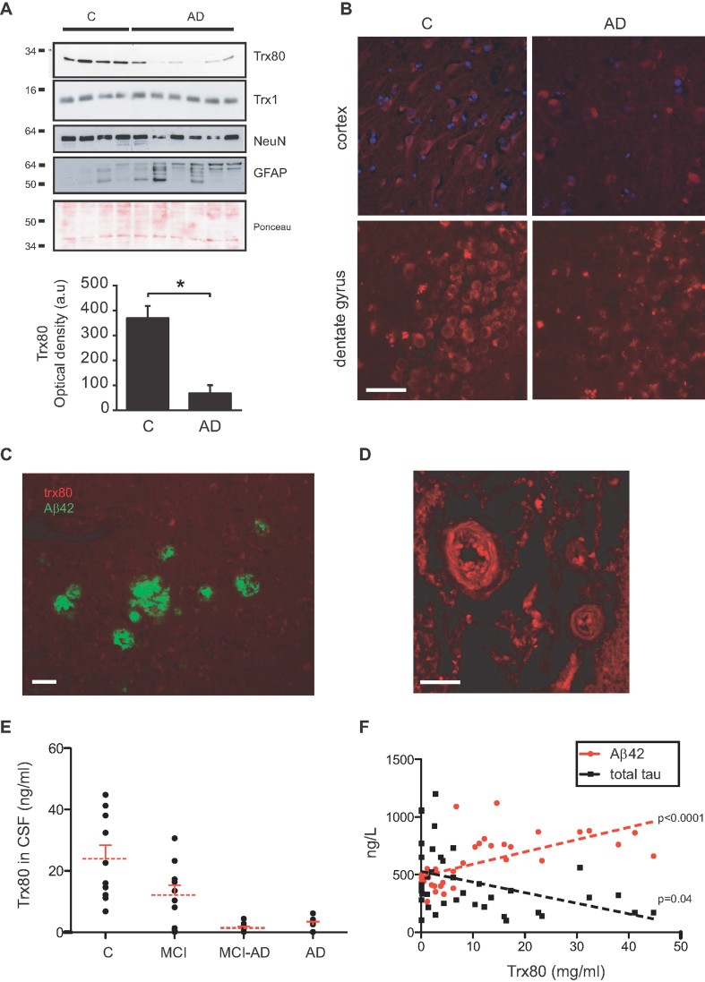
Figure 3. Reduced Trx80 levels in AD.
- Immunoblotting of control and AD brain samples in human brain revealed a significant reduction of Trx80 in AD brains. No changes were found in Trx1 levels. As expected, NeuroN levels were reduced and GFAP levels increased in AD, reflecting neuronal loss and gliosis. Ponceau staining is shown as loading control. Histogram shows data expressed as optical density units and presented as means ± SEM (Mann–Whitney U-test; *p = 0.02).
- Trx80 staining is dramatically decreased in cerebral cortex and dentate gyrus of AD patients.
- Aβ(1–42) staining (green) does not co-localize with Trx80 staining (red) in senile plaques.
- One of the deep vessels with abundant Trx80 staining found in AD brains. Scale bars: 25 µm.
- Levels of Trx80 in CSF, measured by Sandwich ELISA, were reduced in samples from AD and MCI-progressing to AD patients compared to controls and MCI non-progressive patients (ANOVA, Bonferroni's multiple comparison test, p = 0.0001). A significant decrease was also found in non-progressive MCI samples compared to both controls and MCI-AD (p = 0.04).
- Levels of Trx80 in CSF correlated with Aβ42 levels [r = 0.6313; p(two-tailed) = 0.0001] and total tau [r = −0.4070; p(two-tailed) = 0.04].

