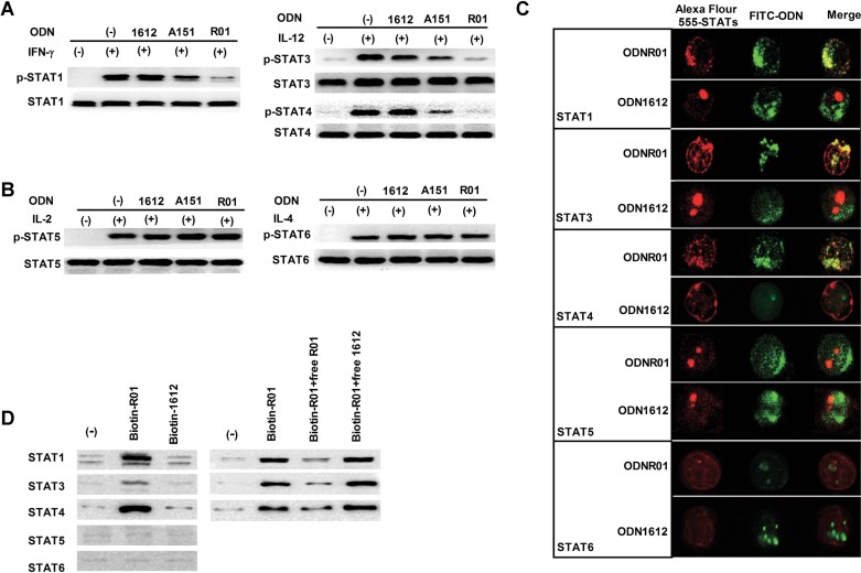
Figure 2. Immunosuppression of STAT1/3/4 phosphorylation by ODNR01 selective binding.
- A,B. STAT and phospho-STAT Western blot analysis of anti-CD3/28 mAb-stimulated CD4+ T cells treated with indicated cytokines, with and without 5 µM of ODNR01, ODNA151 or ODN1612.
- C. Confocal microscopy to detect co-localization of FITC-conjugated ODNs (2.5 µM; green) with different Alex Fluor 555-conjugated anti-STAT rabbit polyclonal antibodies (red) in CD4+ T cells after 24 h of incubation.
- D. Left panels: Immunoblot analysis of STATs from CD4+ T cells pre-incubated with 5 µM biotinylated-ODNs, lysed, and precipitated with avidin agarose beads for STAT detection. Right panels: Immunoblot analysis of STATs from CD4+ T-cell lysates that were pre-incubated with 1 µM biotinylated ODN and 5 µM unlabelled ODN for 1 h and immunoprecipitated with avidin beads for detection of different STAT. The experiments were repeated three times with similar results.

