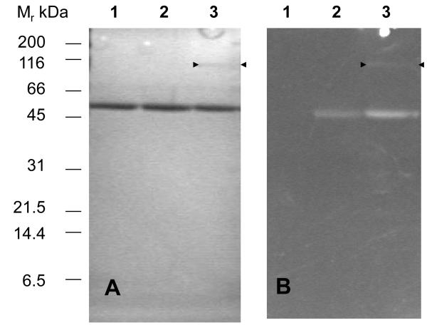
Figure 3. SDS-PAGE of trypanothione reductase inactivated by quinacrine mustard.
TryR (0.1 mg ml−1) was incubated with or without NADPH (150 μM) and QM (20 μM) for 10 min and the reaction quenched by the addition of excess 2-mercaptoethanol (25 mM). Samples were separated by 10% SDS PAGE and examined under UV-illumination followed by staining with Coomassie blue. Panel A: Coomassie stained gel: lane 1, untreated control; lane 2, plus QM without NADPH; lane 3; plus QM with NADPH. The arrow heads point to an additional faint band at 110 kDa. Panel B: UV-illumination, lanes as in panel A.

