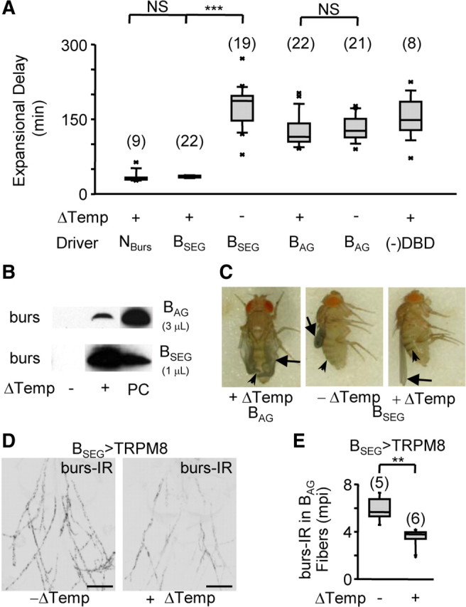
Figure 3.

The BSEG neurons command wing expansion in Drosophila. A, Box plots showing the time required to complete expansional behaviors (i.e., Expansional Delay) for flies expressing TRPM8 in the indicated cell group (Driver) with or without stimulation (Δtemperature). (−)DBD, controls lacking the BursGalDBD-U6A1 hemidriver. As noted in the legend of Figure 1, some flies that attempted expansion after prolonged delays failed to expand their wings, in which case the expansional delay represents the time they took to complete abdominal flexion. The number of flies tested in each group is indicated in parentheses above the bars; the results of t test comparisons between groups are indicated by brackets [NS, not significant (p > 0.05); ***p < 0.001]. B, Western blots showing blood titers of bursicon (i.e., anti-burs immunostaining) resulting from stimulation (Δtemperature) of the BAG (top) or BSEG (bottom). PC, burs-positive control indicates release during normal expansion. Volumes of blood loaded per sample are indicated. C, Wing morphology (arrows) and tanning (arrowheads) of flies expressing TRPM8 in either the BAG or BSEG evaluated 3 h after eclosion. D, Abdominal nerves from BSEG>TRPM8 animals with (right) or without (left) BSEG stimulation immunostained for bursicon using anti-burs antibodies. Scale bars, 50 μm. E, Box plot showing quantified burs-IR data from abdominal nerves of the indicated number of animals of each kind shown in D, demonstrating release of bursicon from these nerves in response to BSEG stimulation. mpi; Mean pixel intensity (background-subtracted). **p < 0.01 by t test. Error bars are SEM.
