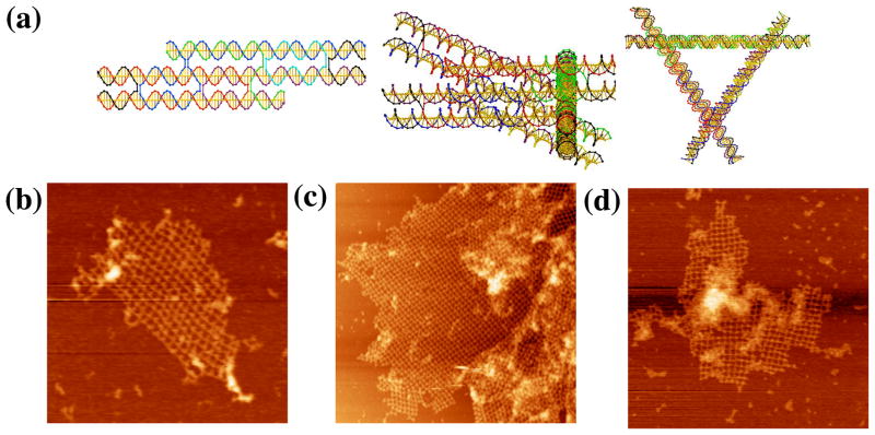
Figure 5. Images of the Skewed TX Triangle. (a) Schematics of the Motif.
The left drawing is the skewed TX structure, and the middle drawing shows a view that emphasizes one of the three sides of the motif. The right view is a 3-fold symmetric picture of the motif. (b–d) The Three Independent Sections Built from Self-Assembly of the Motif. The edges of the three sections are 760, 1700 and 1200 nm, respectively.

