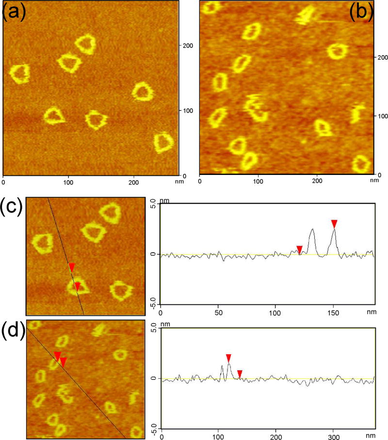
Figure 3.
Atomic Force Micrographs of the PX Triangles. (a) contains images of the triangle, where the frequent appearance of a triangular cavity is evident. (b) shows the blue-green half-triangle; these images are more circular and less triangular than those in (a). (c) contains a cross-sectional analysis of the triangle, and (d) contains a cross-sectional analysis of the half-triangle; note the increased height in (c), relative to (d).

