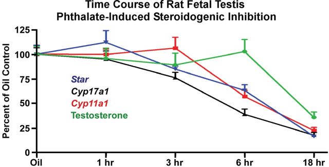
FIG. 1.
Time course of acute phthalate-induced disruption of rat fetal testis steroidogenic gene expression and intratesticular testosterone levels. Sprague Dawley rat dams at GD19 were gavaged with oil vehicle (n = 10) or a single administration of 500mg/kg DBP followed by euthanasia at 1h (n = 5), 3h (n = 5), 6h (n = 5), or 18h (n = 4). Euthanasia for the oil vehicle dams was spread among the 1-, 3-, 6-, and 18-h time points. From fetal testes, steroidogenic gene mRNA levels were quantified by TaqMan-based reverse transcription-polymerase chain reaction relative to Tata binding protein mRNA levels, and intratesticular testosterone levels were measured by radioimmunoassay (Johnson et al., 2011). Shown are the means ± standard errors about the mean. Statistical significance was determined using a one-way analysis of variance and Dunnett’s post hoc test with p < 0.05 considered significant. In this experiment, steroidogenic gene mRNA levels were reduced significantly by 3 (Cyp17a1) or 6 h (Cyp11a1 and Star) postexposure, but significant intratesticular testosterone reductions were not observed until 18h postexposure. Expression of steroidogenic genes remained significantly lower through the 18-h time point. Thus, inhibition of steroidogenic pathway gene expression preceded reductions in intratesticular testosterone levels.

