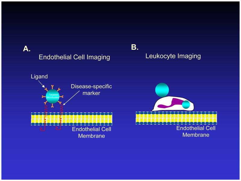
Figure 1.
Schematic diagram of vascular endothelial lining and approaches for ultrasound molecular imaging in which ultrasound contrast agents, or microbubbles, adhere to endothelium. A. Microbubbles with a targeting ligand on their surface can bind specifically to a molecule overexpressed by endothelium in cardiovascular disease, such as a leukocyte adhesion molecule. B. Activated leukocytes may bind or phagocytose microbubbles, which remain acoustically active for a brief period of time, allowing ultrasound detection of leukocytes which have adhered to inflammatory endothelium. Figure not drawn to scale.

