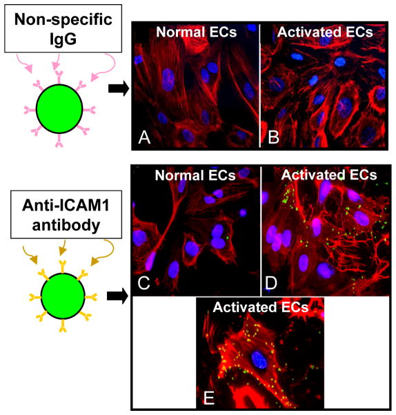
Figure 2.
In vitro studies demonstrating proof of the concept that microbubbles can adhere to a biological surface via a specific targeting ligand. Fluorescent microbubbles (green) were conjugated to either non-specific IgG (control) or to antibody directed against intercellular adhesion molecule 1 (ICAM1) and allowed dwell with cultured human coronary artery endothelial cells (ECs, F-actin labeled with rhodamine, red). Control microbubbles did not adhere to cells at baseline (A) or after endothelial activation to overexpress ICAM1 (B). Anti-ICAM1 microbubbles adhered minimally to non-activated cells due to constitutive expression of ICAM1 (C), and adhered abundantly to activated endothelial cells (D). Panel E is a higher power image of a single activated cell to which multiple ICAM1-targeted microbubbles have adhered. Reprinted with permission from Ref 22

