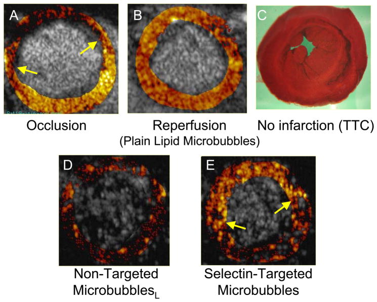
Figure 4.
Ischemic memory imaging of myocardium using microbubbles targeted to bind to P-selectin in a rat model of 15 minute coronary occlusion/reperfusion. Short axis ultrasound images of left ventricular myocardium at mid-papillary muscle level are background subtracted, color-coded frames in which shades of red, progressing to orange, yellow, and white, indicate increasing contrast change. A. During coronary occlusion, there is a contrast defect corresponding to the risk area (region between arrows). B. After release of the coronary occlusion, myocardial contrast echo perfusion imaging with non-targeted lipid bubbles confirms reperfusion to the anterior wall. C. Post-mortem staining with triphenyl tetrazolium chloride (TTC) shows no infarction. D. During reperfusion, imaging after intravenous injection of non-targeted lipid bubbles bearing sialyl Lewisc as the control ligand, shows no evidence of persistent myocardial contrast enhancement. E. During reperfusion, imaging after intravenous injection of microbubbles targeted to P-selectin via sialyl Lewisx demonstrates persistent contrast enhancement in the area that was previously ischemic. Reprinted with permission from Ref 27.

