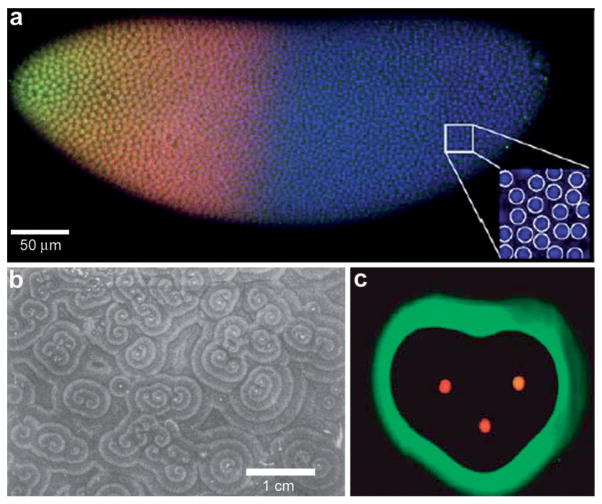
Fig. 3.
Examples of chemical patterning at the multi-cellular level. (a) Scanning confocal microscope image of a Drosophila embryo in early nuclear cycle, stained for DNA (blue), Hb (red), and Bcd (green); scale bar 50 μm. Inset (28 × 28 μm2) shows how DNA staining allows for automatic detection of nuclei. (Reprinted with permision from ref. 30. Copyright (2007) by Elsevier.) (b) Organized waves of cell movement during aggregation in Dictyostelium discoideum. (Reprinted with permission from ref. 33. Copyright (1981) by The American Association for the Advancement of Science). (c) Experimental result showing heart-shaped GFP patterns formed based on the placement and initial concentrations of sender cells (three sender disks are shown in the image in red color) expressing DsRed-Express. (Reprinted with permission from ref. 35. Copyright (2005) by The Nature Publishing Group).

