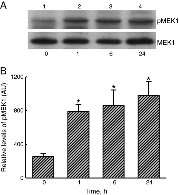
Figure 6.
Effects of lipoteichoic acid (LTA) on the phosphorylation of mitogen-activated/extracellular signal-regulated kinase kinase (MEK)1. A549 cells were exposed to 30 μg/ml LTA for 1, 6, and 24 h. Phosphorylated MEK1 (p-MEK1) was immunodetected (A, top panel). ERK2 was detected as the internal standard (bottom panel). These immunorelated protein bands were quantified and analyzed (B). Each value represents the mean ± SEM for n = 6. An asterisk (*) indicates that a value significantly differed from the control group, p < 0.05. AU, arbitrary unit.

