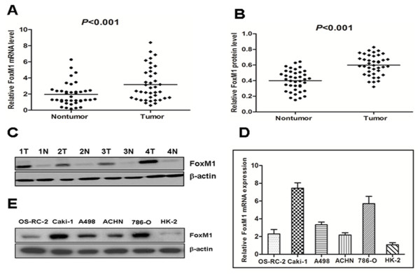
Figure 1.
The expression of FoxM1 mRNA and protein in the human ccRCC surgical specimens and RCC cell lines, as evaluated by real-time quantitative PCR and western blot.A, The relative mRNA expression of FoxM1 was higher in 39 ccRCC tumor tissues than in matched adjacent nontumorous tissues (P < 0.001). B, The FoxM1 protein expression was higher in the tumor tissues than in matched adjacent nontumorous tissues (P < 0.001). C, Expression of FoxM1 protein in four representative pairs of ccRCC tissues is presented. N, nontumorous tissues; T, ccRCC tissues. D, The FoxM1 mRNA expression in human RCC cell lines was higher in the OS-RC-2, Caki-1, A498, ACHN and 786-O cells, particularly in the Caki-1 and 786-O cells, compared with the normal proximal tubule epithelial cell line HK-2. E, The FoxM1 protein expression was elevated in the OS-RC-2, Caki-1, A498, ACHN and 786-O cells compared to the normal proximal tubule epithelial cell line HK-2.

