Abstract
From a genetic screen for Drosophila melanogaster mutants with altered ethanol tolerance, we identified intolerant (intol), a novel allele of discs large 1 (dlg1). Dlg1 encodes Discs Large 1, a MAGUK (Membrane Associated Guanylate Kinase) family member that is the highly conserved homolog of mammalian PSD-95 and SAP97. The intol mutation disrupted specifically the expression of DlgS97, a SAP97 homolog, and one of two major protein isoforms encoded by dlg1 via alternative splicing. Expression of the major isoform, DlgA, a PSD-95 homolog, appeared unaffected. Ethanol tolerance in the intol mutant could be partially restored by transgenic expression of DlgS97, but not DlgA, in specific neurons of the fly’s brain. Based on co-immunoprecipitation, DlgS97 forms a complex with N-methyl-D-aspartate (NMDA) receptors, a known target of ethanol. Consistent with these observations, flies expressing reduced levels of the essential NMDA receptor subunit dNR1 also showed reduced ethanol tolerance, as did mutants in the gene calcium/calmodulin-dependent protein kinase (caki), encoding the fly homolog of mammalian CASK, a known binding partner of DlgS97. Lastly, mice in which SAP97, the mammalian homolog of DlgS97, was conditionally deleted in adults failed to develop rapid tolerance to ethanol’s sedative/hypnotic effects. We propose that DlgS97/SAP97 plays an important and conserved role in the development of tolerance to ethanol via NMDA receptor-mediated synaptic plasticity.
Introduction
In humans and other organisms, repeated consumption of alcohol leads to the development of tolerance, defined as an acquired resistance to the physiological and behavioral response to a particular concentration of alcohol (reviewed in [1], [2]). The development of tolerance is a complex event associated with multiple physiological as well as functional changes. Yet, our knowledge of the mechanisms by which prolonged or repeated ethanol exposure exerts its effects in the brain to alter behavior remains limited.
The fruitfly Drosophila melanogaster has been developed as a useful model system to identify molecules and pathways involved in the development of ethanol tolerance [3], [4], [5], [6], [7], [8], [9], [10], [11], [12], [13], [14], [15]. We identified a mutant, which we named intolerant (intol) that exhibits greatly reduced ability to develop tolerance. The intol mutant was found to carry a mutation in the gene discs large 1 (dlg1). The protein products of dlg1, Discs Large A (DlgA) and DlgS97, are the highly conserved fly homologs of the mammalian PSD-95 and SAP97 scaffolding proteins, respectively. They are involved in targeting and clustering membrane receptors, including a known ethanol target, the N-methyl-D-aspartate receptor (NMDAR), at the synapses [16].
Acute alcohol exposure antagonizes NMDAR function, while chronic alcohol consumption leads to an increase in NMDAR-mediated neurotransmission [17]. The subsequent alterations in synaptic function are hypothesized to involve changes in receptor density and phosphorylation, which are thought to impact receptor clustering and/or complex formation [18], [19], [20]. The resulting changes in glutamate receptor signaling at the synapse have been implicated in the development of ethanol dependence, tolerance, and addiction [1], [21]. Therefore, proteins that regulate these synaptic remodeling events are thought to gate the development of ethanol-induced synaptic and behavioral plasticity [1], [17].
Via alternative splicing, the Drosophila dlg1 locus encodes two major protein products, DlgA and DlgS97, which exhibit largely similar domain structures [22], [23]. Interestingly, the mammalian isoforms of these proteins, PSD-95 and SAP97, respectively, are encoded by two separate genes [24]. Both DlgA and DlgS97 are expressed at larval neuromuscular synapses, where DlgA is important for normal development and the organization of an intricate protein network in the postsynaptic region [25]. Recently, DlgA and DlgS97 were shown to be differentially expressed during development and adulthood: only DlgA is required for adult viability, while specific loss of DlgS97 leads to perturbation of circadian activity and courtship [26]. The mammalian homolog of DlgS97, SAP97, is ubiquitously expressed in the brain and can localize to pre- and/or post-synaptic sites of excitatory or inhibitory synapses [27]. SAP97 has been shown to interact with the C-terminus of NMDA and α-amino-3-hydroxy-5-methyl-4-isoxazolepropionic acid (AMPA) receptors [28], [29], [30], [31]. Recently, SAP97 has also been implicated in trafficking of NMDARs at the cell surface by sorting them through an unconventional secretory pathway [32].
In this study, we report a novel role for DlgS97 and SAP97 in the development of tolerance to ethanol. By testing multiple independently isolated dlg1 alleles, we determined that DlgS97 is required for the development of rapid ethanol tolerance in Drosophila. We further demonstrate that hypomorphic mutants of Nmdar1, encoding the Drosophila NMDA receptor 1 (dNR1) subunit, or Camguk/Caki, encoding the homolog of CASK that interacts with the L27 domain of DlgS97 [22], exhibit reduced ethanol tolerance. Co-immunoprecipitation of dNR1 and DlgS97 confirmed a physical interaction of DlgS97 and NMDARs. We propose that this interaction is of functional relevance to ethanol tolerance development. Lastly, we show that adult mice with reduced expression of SAP97 display deficits in the development of rapid tolerance to the sedative/hypnotic effects of ethanol, measured using regain of the loss of righting response (LORR) [33], [34], [35].
Materials and Methods
Ethics Statement
All animal protocols were approved by the Ernest Gallo Clinic and Research Center (EGCRC) Institutional Animal Care and Use Committee (approval number 10.01.205).
Drosophila Strains and Genetic Analysis
All flies were raised and maintained on standard cornmeal molasses agar at 25°C and 70% humidity. For the behavioral screen, P-element insertion lines were generated by mobilizing the pGawB transposable element [36], [37]. The tolerance screen that led to the identification of intolerant (GenBank accession number: JM426603) screened a small number of strains, 42 lines, comprising a subset of this P-element insertion collection. The control line for the intolerant mutant was the otherwise isogenic, parental background strain, w;iso (CJ1) (2202U, [38]). For all behavioral experiments, fly lines were outcrossed for five generations to 2202U flies prior to behavioral testing. Fly strains harboring mutant alleles of dlg1 were obtained from the following sources: dlg1m52 [39]; Bloomington Stock Center); dlg15779 (Bloomington Stock Center); dlgNP4134, dlgNP768, dlgN7P229, and dlgNP1102 (GAL4 Enhancer Trap Insertion Database (GETDB)). The following dlg1 constructs were utilized: UAS-eGFP-dlgA [40] and UAS-eGFP-DlgS97 [41], which were generously provided by Dr. Uli Thomas at the Leibniz Institute for Neurobiology in Germany. The GAL4c747 enhancer stock and fly stocks harboring NMDAR1 mutant alleles, NR1EP331 and NR1EP3511 [42] were obtained from the Bloomington Drosophila Stock Center. The Nmdar1 deficiency stock, NR190B.0 (in which the itpR83D IP3 receptor gene is also deleted) was generously provided by the laboratory of Dr. Hasan at the National Centre for Biological Sciences, India [43]. The GFP-balanced stock (90B.0/TM3-Ser-GFP) was generated by crossing homozygotic deficiency stock with GFP balancer flies (TM6b/TM3-Ser-GFP).
The P-element insertion in the dlg1-intol mutant was characterized using inverse PCR and DNA sequencing. Mutants of Drosophila CASK (Camguk/Caki) were generated by imprecise P-element excision and have been described [44].
Ethanol Behavioral Assays
Unless otherwise stated, adult (2–4 days post-eclosion) male flies were used for all experiments. Flies were collected under CO2 anesthesia 2–3 days before experiments. Ethanol sensitivity and tolerance were quantified using the inebriometer, an apparatus that measures the loss of postural control with increasing time of exposure to ethanol vapor of a sample population of flies [4], [6], [45], [46]. To quantify ethanol tolerance, a sample of ∼120 flies was exposed to ethanol vapor at a relative flow rate of 60/40 ethanol vapor/humidified air for 30 min, transferred to large vials containing fly food, and allowed to recover from sedation for an additional 3.5 hr (total time of 4 hr elapsed between the first and the second exposures). In parallel, a second, control sample otherwise identically treated was exposed to humidified air without ethanol at an equivalent total flow rate and allowed to recover. Following recovery, each pair of ethanol pre-exposed and control samples were assayed using side-by-side inebriometers. Tolerance was calculated in minutes as the difference in inebriometer mean elution time (MET) between naive and ethanol-pre-exposed flies using the formula (METexp2-METexp1) [7].
For some experiments, where stated, sensitivity and tolerance to ethanol were measured using sedation assays, performed essentially as previously described [37]. Sample populations of ∼25–30 flies were pre-exposed to ethanol vapor or to humidified air (control) as described above, and 4 hr later were tested for loss of ability to stand upright during exposure to ethanol vapor in the Booz-O-Mat apparatus. Sedation sensitivity was quantified as the ST50, the time for 50% of the flies in a sample population of 25–30 flies to become sedated, and tolerance was calculated as the difference in ST50 between flies previously exposed to ethanol vs. control (ST50 exp2-ST50 exp1). Chronic tolerance, ethanol sedation assay and ethanol absorption and metabolism are described in supplemental methods.
Antibodies and Immunofluorescence Microscopy
For larval NMJ staining, wandering third instar larvae were dissected in Ca2+-free phosphate-buffered saline (PBS), fixed in 4% paraformaldehyde in PBS for 20 min, washed several times with PBS containing 0.1% Triton X-100 (PBST), and blocked in PBST supplemented with 5% fetal bovine serum (FBS) and 0.1% bovine serum albumin (BSA, blocking buffer) for 1 hour at RT. Following blocking, larvae were incubated with primary antibodies in blocking buffer at 4°C overnight. For adult brain staining, flies were collected 1–3 days after eclosion. CNSs were dissected in PBS, fixed in 4% paraformaldehyde in PBS for 20 min at room temperature, washed three times for 10 mins in PBS containing Triton X-100 at 0.3%, blocked for 1 hour at RT in blocking buffer and incubated with primary antibodies in blocking buffer overnight at 4°C. Samples were washed three times for 10 mins in PBST at RT, and secondary antibodies were applied in blocking buffer for 2 hr at RT. After washing three times for 10 min in PBST and once in PBS, samples were mounted in Vectashield (Vector Labs, Burlingame, CA). The following primary antibodies were used: mouse anti-DLGPDZ (1∶2000, [40]) Guinea-pig anti- DlgS97N (1∶4000, the polyclonal anti- DlgS97N antibodies were generated by immunizing both rabbits and guinea-pigs with the amino-terminal 205 amino acids of dSAP97 fused to GST (Cocalico Biologicals, Inc., Reamstown, PA), rabbit anti-NR1 antibody (used at 1∶300 for the western and was generated by injection of C-terminal peptide stretch containing KTRPQQSVLPPRYSPGYTSD; ZyMed Laboratories, Inc.), mouse anti FASII antibody (1∶500, 1D4 from Developmental Studies Hybridoma Bank), mouse anti GFP antibody (1∶500, Molecular Probe) and Cy3-conjugated Goat anti-horseradish peroxidase (1∶250, Jackson ImmunoResearch). Fluorescently-labeled secondary antibodies were purchased from Molecular Probes (1∶500, Alexa-495, −568 and −650). Larval and brain images were captured using a Leica TCS SP2 confocal microscope.
Immunoblotting and Immunoprecipitation
For protein analysis of mutant alleles, 3 larval body wall muscles or 10 adult heads per sample were homogenized in 50 µl of SDS sample buffer (InVitrogen LDS 4X buffer containing 50 mM DTT), separated by SDS-PAGE and blotted onto nitrocellulose transfer membrane. After blocking in Tris buffered saline containing 0.1% Tween (TBST) and 5% dry milk, the membrane was incubated with anti-DlgPDZ at 1∶500, anti-DlgS97N antibody at 1∶1000, or mouse anti-Tubulin antibody (Sigma, St. Louis, MO) at 1∶10,000. For immunoprecipitation of protein complexes from adult head extracts, flies were collected 2–5 days after eclosion and frozen at –80°C for at least a day. Approximately 100 µl volume of adult heads were collected via sieve and homogenized in 1 ml of RIPA buffer containing 50 mM Tris-HCl, pH7.5, 150 mM NaCl, 1 mM EDTA, 1% Nonidet P-40, 0.5% sodium deoxycholate, 0.1% SDS and protease inhibitor cocktail (purchased from Roche). Solubilization was carried out at 4°C for 60 min, followed by centrifugation at 100,000×g for 60 min at 4°C. Another round of re-solubilization was performed on the pellet after the centrifugation. The supernatant was collected, combined and reconstituted in a final volume of 4 ml of RIPA buffer with final concentration of 0.1% Triton X-100. Subsequently, this protein extract was dialyzed for 16 hr in 50 mM Tris, pH 7.5, containing 0.1% Triton X-100 at 4°C. For immunoprecipitation, 2 µl of appropriate antiserum was added to 50 µl (resuspended resin) of protein A beads (GE Healthcare) in PBT (PBS containing 0.1% Triton X-100) for at least 2 hrs at 4°C. Protein A beads were then pelleted, washed in PBT, incubated with 1 ml of the detergent-solubilized protein extract for 4 hrs at 4°C with constant rotation. The beads were then washed with 50 mM Tris, pH 7.5, containing 0.1% Triton X-100 and 150 mM NaCl. After the wash, the beads were resuspended in 4X SDS sample buffer (40 µl), heated for 15 min at 80°C, resolved by SDS-PAGE (7% gel) and transferred to a nitrocellulose membrane. Following blocking in TBST/5% dry milk, the membrane was washed in PBS and incubated in primary antibody (rabbit polyclonal anti-NR1 peptide antibody, diluted 1∶1,000) in blocking solution (TBS with 5% BSA) overnight at 4°C.
For immunoblotting mouse brain tissue, brains were isolated from mice 3–4 weeks after the final injection of Tamoxifen and homogenized in 500 µl of RIPA buffer containing protease inhibitors and incubated on ice for 1 hour. Protein estimation was performed using the BCA reagent (Pierce, Rockford, Illinois). 20–30 µg of protein was electrophoresed and transferred on to a 0.45µm PVDF membrane (Invitrogen, Carlsbad, CA). Monoclonal antibody against Dlg was from BD transduction laboratories (CA, USA). Monoclonal antibody against Actin was from Sigma (St. Louis, MO).
Floxed Sap97 Mice
Floxed Sap97 mice were generated and maintained on a mixed C57B6J/129J mice in Dr. Richard Huganir’s laboratory [47] at Johns Hopkins University. For tolerance experiments, these mice were backcrossed to C57B6/J mice for 3 generations. Homozygous floxed mice were then crossed to mice carrying the ESR-Cre transgene on a C57B6J background. Heterozygous Sap97fl/+;cre mice were then crossed with Sap97fl/fl mice to generate the Sap97fl/fl, Sap97fl/+;cre and Sap97fl/fl;cre littermates used in this study. 8-week old mice were injected with 120–130 mg/kg Tamoxifen (TM, Sigma, St. Louis, MO) dissolved in 10% (v/v) ethanol solution in corn oil (Sigma, St. Louis, MO) for 4 days. Ethanol volume per injection was less than 10 µl and the last injection was given at least 3 weeks before measuring ethanol LORR.
Rapid Ethanol Tolerance in Mice
Rapid tolerance to the sedative/hypnotic effects of ethanol was measured using LORR assay. 3–4 weeks after the last TM injection, male mice were injected with 4 g/kg ethanol (20% w/v in saline) on day 1. After ethanol injection, mice were placed on their backs. Mice were determined to have lost their righting reflex when they were unable to right themselves three times within 30 seconds. The animals were deemed to have regained their righting response when they were able to right themselves three times in 30 seconds. LORR duration was calculated as the time interval between when the righting response was lost and when it was regained. The procedure was repeated on Day 2 and duration of LORR was recorded as on Day 1. Tolerance was calculated as the difference in the duration of LORR on Day 1 and Day 2.
Quantitative PCR
RNA was extracted from the brains of 8–12 week old male and female Sap97fl/fl, Sap97fl/+; cre , and Sap97fl/fl;cre mice 3–4 weeks after TM injection. Complementary DNA (cDNA) was generated from 1 µg of RNA using reagents from Applied Biosystems (Foster City, CA, USA). Following synthesis, cDNA was diluted 1∶10 in water. TaqMan QPCR was performed using standard thermal cycling conditions using an ABI PRISM 7900 Sequence Detection System (Applied Biosystems). Amplification reaction contained 5 µl of cDNA template, 1x PCR Master Mix, 100 nM each of forward and reverse primers and 200 nM of FAM-labeled probe in a final volume of 10 µl. Sap97 primer/probe set spanning the boundary of exon 10 and 11 of the mouse Dlg1 gene was obtained from Applied Biosystems (Assay ID: Mm01344472_m1).
Ethanol Metabolism
One week after the LORR assay, mice were injected with 4 g/kg ethanol and tail blood samples were obtained at 30, 60, 90, 120, 180 minutes post injection. Blood samples were stored at −80°C until BEC levels were measured using an NAD-ADH enzymatic assay [48].
Results
Identification of intolerant, a Drosophila Mutant with Reduced Ethanol Tolerance
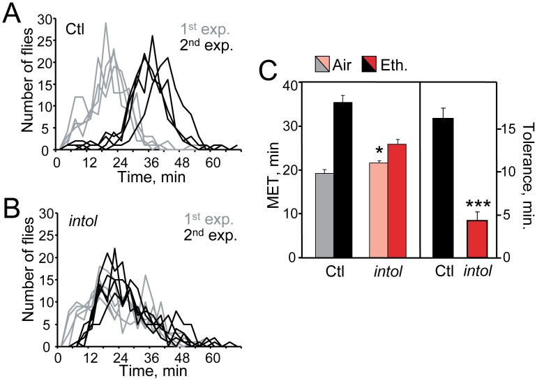
During exposure to ethanol vapor, adult flies become hyperactive, uncoordinated, and eventually sedated [49], [50]. The behavioral effects of ethanol include loss of postural control, which may readily be quantified in the inebriometer, a vertical column containing evenly spaced mesh platforms in which ∼120 flies per sample are exposed to ethanol vapor and counted as they fall from the column over time [45], [46]. Under our standard assay conditions, ethanol-naïve control flies emerged from the inebriometer with a mean elution time (MET) of ∼20 min (Fig. 1A, 1C). As previously reported, a single exposure to ethanol vapor that is sufficient to sedate flies (30 minutes at a 60/40 ratio of ethanol vapor/humidified air) leads to the development of tolerance, manifested as an increase in inebriometer MET measured 4 hours after the initial exposure, a time at which the first dose has been entirely metabolized (control flies are pre-exposed to humidified air only [4], [6]). This acquired resistance, or tolerance, correlates with an increase in the absorbed ethanol levels needed to induce loss of postural control, and has been quantified as the difference in MET between the first and second ethanol exposures [4], [7] (Fig. 1C).
Figure 1. The intol mutant exhibits drastically reduced ethanol tolerance development.
(A) Normal ethanol tolerance is displayed by control flies (parental strain, Ctl). Ethanol-naïve control flies eluted from the inebriometer with a mean elution time (MET) of 19.2±0.9 min (1st exposure, gray), while the elution profiles of Ctl flies that had been exposed to ethanol 4 hr earlier, were strongly shifted to the right (2nd exp., black; MET = 35.4±1.6 min). (B) Ethanol pre-exposure under otherwise identical conditions did not produce a similar increase in resistance in the intol mutant (1st exp., gray, MET = 21.6±0.5 min; 2nd exp., black, MET = 25.9±1.1 min). Inebriometer elution profiles for Ctl (A) and intol mutant flies (B) are shown; n = 4 (Ctl) or 5 (intol). Here and elsewhere, error bars represent standard error of the mean (SEM), and n refers to the number of samples, not the number of flies. (C) MET and tolerance values for Ctl and intol flies for inebriometer data represented in panels (A) and (B). The intol mutant displayed strongly reduced tolerance of 4.3±1.0 min vs. 16.2±1.2 min for Ctl flies (***, [F(1,7) = 55.53, p<0.001], one-way ANOVA). Additionally, ethanol resistance in the intol mutant showed a modest but significant increase, with MET = 21.6±0.5 min vs. 19.2±0.9 min for Ctl flies (*, [F(1,7) = 6.511, p<0.05]).
To identify molecules and pathways involved in ethanol tolerance development, we carried out a small-scale genetic screen for Drosophila mutants exhibiting an altered response to repeated ethanol exposure. Here we report the identification of one mutant, intolerant (intol) that showed drastically reduced tolerance development compared to the parental strain (Fig. 1B, 1C). The intol mutant also exhibited a small but statistically significant decrease in ethanol sensitivity (Fig. 1C, left panel). Ethanol absorption and metabolism of the intol mutant appeared unaffected (Methods S1, Fig. S1).
Ethanol tolerance in Drosophila has been previously shown to be proportional to the ethanol concentration and time of pre-exposure, and requires a threshold dose [4]. We found that longer ethanol pre-exposures increased tolerance of control flies, as expected, but failed to induce tolerance in the intol mutant (Methods S1, Fig. S2). The intol tolerance defect appeared recessive, as heterozygous mutant flies developed normal tolerance (Fig. S3). The intol mutant was further characterized for development of chronic tolerance, a mechanistically distinct form of tolerance produced by long-term, low-level ethanol exposure [6]. In this paradigm, intol also exhibited reduced tolerance (Fig. S4).
The intol Mutation Disrupts Expression of DlgS97, a Neuronal Isoform of Dlg1
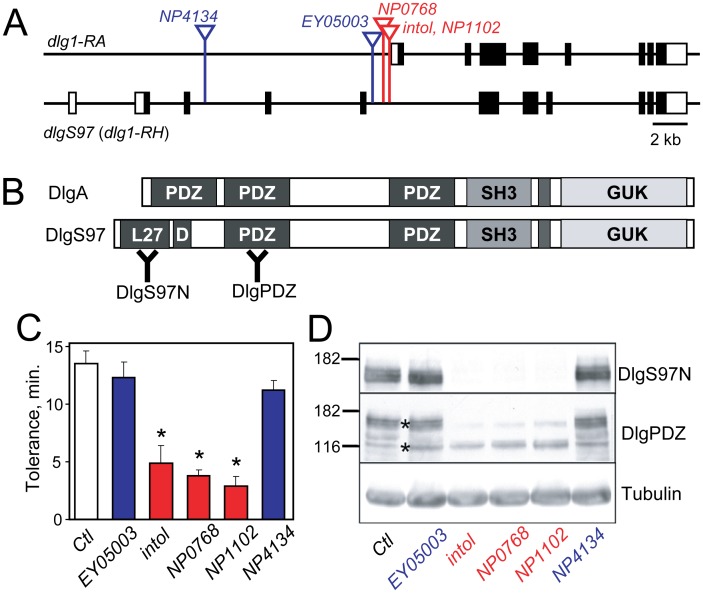
Inverse PCR and DNA sequence analysis revealed that the P element in the intol mutant was inserted within an intron of the gene discs large 1 (dlg1) (Fig. 2A). To further investigate the involvement of dlg1 in ethanol tolerance, we characterized the effects of additional, independently identified P element insertions in the dlg1 locus (Fig. 2A). Ethanol tolerance was quantified for 4 additional potential dlg1 mutants (Fig. 2C). Of these, two insertions, NP1102 and NP0768, exhibited tolerance defects similar to intol, implying that the tolerance defects seen in these mutants is caused by the P element insertion in dlg1. Two other insertions, EY05003 and NP4134, produced no significant decrease in tolerance compared to control flies (Fig. 2C). Similar results were obtained for these mutants in the chronic tolerance exposure paradigm (Fig. S4).
Figure 2. Genomic structure, additional mutants and protein products of the dlg1 locus.
(A) Schematic representation of dlg1 genomic structure for two major characterized transcripts, dlg1-RA and dlg1-RH. Exons are represented with boxes, protein-coding sequences by filled boxes, and introns by lines. Vertical lines with inverted triangles denote independent P-element insertions in dlg1 including intol. (B) Schematic depiction of DlgA (dlg1-RA product) and DlgS97 (dlg1-RH product) showing characterized protein domains. Two distinct regions used for generating antibodies are indicated as “Y” in bold. (C) Two additional transposon mutants in dlg1, NP0768 and NP1102, whose positions are shown in red in panel (A), also showed reduced ethanol tolerance (***, [F(5,18) = 19.973, p<0.001], one way ANOVA followed by Holm-Sidak test, n = 4). (D) The same subset of dlg1 mutants showed loss of DlgS97. Immunoblot analysis was performed on protein from adult heads from control flies and the dlg1 mutants EY05003, intol, NP0768, NP1102, and NP4314 using pan-Dlg (“DlgPDZ”) and DlgS97-specific antibodies; tubulin was used as a loading control.
We next addressed the effect of these P element insertions on Dlg protein expression. The dlg1 locus is complex and encodes several transcripts through use of multiple transcriptional start sites and alternative splicing (Flybase, http://flybase.org; diagrammed in simplified form in Fig. 2A and 2B). Since dlg1 null mutations cause lethality [39], we hypothesized that the P-element mutations affecting ethanol tolerance might decrease expression of a subset of dlg1 products. Transcripts encoded by dlg1 are known to produce two major functional proteins, DlgA and DlgS97 (Fig. 2B), whose expression has been well characterized at the larval neuromuscular junction (NMJ) [22], [23]. DlgS97, whose protein sequence is largely shared with DlgA but contains an additional N-terminal L27 (lin-2/lin-7) heterodimerization domain [51] not present in DlgA, was shown to be the predominant form expressed in adult brain, and was implicated in circadian rhythms, phototaxis, and courtship [26]. To investigate how dlg1 expression might relate to the ethanol tolerance defects of the mutants, protein extracts from heads of adult dlg1 mutant and control flies were compared by western analysis using two different antibodies: one, anti-DlgPDZ, directed against a PDZ domain and thus recognizing DlgA and DlgS97 [40], and the second, anti-DlgS97N, specific for DlgS97 isoforms [41] (Fig. 2D). In protein isolated from adult fly heads, expression of DlgS97 was undetectable in the 3 mutants (intol, NP0768 and NP1102) that displayed reduced ethanol tolerance. DlgS97 levels were unaffected in the two other mutants with normal tolerance, EY05003 and NP4134. At the larval NMJ, qualitatively similar results were obtained, with DlgS97 being undetectable in the intol, NP0768 and NP1102 mutants (Fig. S5). These results show that a subset of P element insertions, including that in the intol mutant, disrupt expression specifically of DlgS97, and that this dlg1-encoded isoform is most likely required for the development of normal ethanol tolerance.
DlgS97 Expression in the Adult Brain
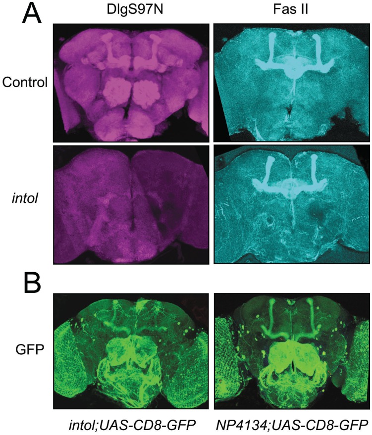
Although the expression pattern and function of dlg1 at the larval neuromuscular junction have been well elucidated, its role in the adult CNS has not been widely studied. We first determined the expression pattern of both dlg1-encoded isoforms, DlgA and DlgS97, in the adult brain. In wild-type adults, DlgS97 protein was prominently expressed in the mushroom body and antennal lobes (Fig. 3A), regions that are important for olfactory learning and memory [52]. This expression was abolished in intol mutant adult brains (Fig. 3A). In the same preparation, double-labeling with the DlgPDZ antibody (recognizing both Dlg isoforms) resulted in staining identical to that seen with the DlgS97N antibody, consistent with the finding that DlgS97 is the predominant form expressed in the adult brain [26]. Co-immunolabeling with FasII, a molecule that normally localizes to the mushroom body and central complex, revealed no gross anatomical defects in the brains of intol mutant flies (Fig. 3A, right panels).
Figure 3. The intol mutant shows loss of DlgS97.
(A) Whole-mount brains of Control (upper panels) and dlg1intol (lower panels) adult flies stained with anti-DlgS97 antibody (left panels, red) and anti-FASII antibody (right panels, blue). (B) Whole-mount brains of adult flies expressing UAS-CD8-GFP driven by dlg4134GAL4 or dlg16–99GAL4 and stained with anti-GFP antibody. Each image represents a stack of 20–25 optical sections taken at 0.5 µm steps.
The P element present in the dlg mutant alleles, including intol flies, contains the coding region for the yeast transcription factor GAL4, positioned such that GAL4 will be expressed under the control of local genomic promoters/enhancers, likely those that normally regulate Dlg expression. By crossing intol flies to flies carrying a UAS-GFP transgene, in which Green Fluorescent Protein (GFP) is expressed via GAL4-responsive UAS promoter elements, dlg enhancer-directed expression of GAL4 can be visualized. Using this method, we analyzed the potential endogenous Dlg expression pattern using two independent dlg alleles with different P element insertion sites. In NP4134, an allele that showed wild-type levels of DlgS97 protein expression and normal ethanol tolerance, GFP appeared expressed moderately in the mushroom body and strongly in the antennal lobes, subesophageal ganglion, and some additional identifiable neurons, such as the dorsal giant interneurons (DGI), neurons of the pars intercerebralis (PI) and lateral neurons (Fig. 3B, right panel). Flies in which GFP was driven by the GAL4 element of the intol insertion showed a GFP expression pattern similar to that seen in NP4134;UAS-GFP flies. However, there were some clear differences; in particular, there was a consistent reduction in GFP staining in the mushroom body, suggesting that the GAL4 element in intol only partially recapitulates the wild-type dlg1 expression pattern in the adult fly brain (Fig. 3B, left panel). This has implications for experiments performed using UAS-dlgA and UAS-dlgS97 transgenes to assess behavioral rescue as described below.
Both DlgA and DlgS97 are highly expressed in the post-synaptic regions of type I boutons at the NMJ ([53] and our unpublished observations). Accordingly, we examined 3rd instar larval NMJs to test whether expression of DlgS97 was lost in the dlg1 mutant larvae. In 2 different tolerance-defective mutants, intol and NP1102, DlgS97 staining appeared completely absent, whereas synaptic accumulation of DlgA persisted (Fig. S5). As a control, we examined staining in a dlg1 null (lethal) mutant, dlgm52, at the larval NMJ and found that expression of both DlgA and DlgS97 was no longer detectable (Supplementary Fig. S5). Despite the lack of DlgS97 at the larval NMJ, electrophysiological analysis of intol revealed no impairment of synaptic transmission: excitatory postsynaptic potential (EPSP) amplitude, average amplitude of spontaneous miniature release events (mEPSPs), and presynaptic release, as estimated by the average EPSP/average mEPSP were normal (G. Davis, unpublished observations). These data are consistent with temporally distinct requirements for DlgA, needed for normal development, and DlgS97, needed for normal adult behavior.
Rescue of intol Ethanol Tolerance Phenotype by Mushroom Body Expression of DlgS97
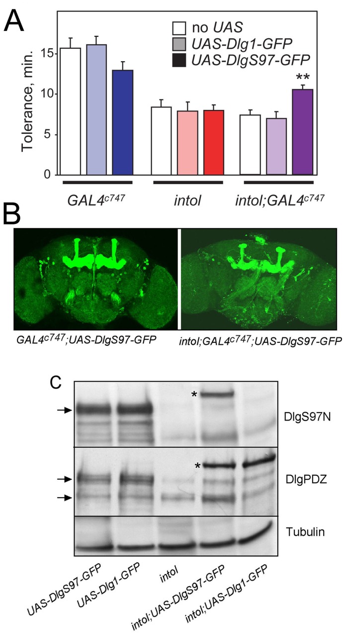
To further investigate the involvement of DlgS97 in ethanol tolerance, we performed behavioral rescue experiments utilizing two different dlg transgenes. We generated wild-type and intol mutant flies harboring either UAS-DlgA-GFP or UAS-DlgS97-GFP. In the intol mutant, the transposon-encoded GAL4 appeared to direct expression of the UAS construct in a similar but not identical pattern to endogenous DlgS97, described above (Fig. 3). Expression of either DlgA or DlgS97 in the pattern dictated by GAL4 expression in intol flies did not rescue the defective ethanol tolerance of the mutant (Fig. 4A, middle bars). We verified transgene expression of DlgS97N and DlgA protein by western analysis (Fig. 4C). Both UAS-DlgA-GFP and UAS-DlgS97-GFP transgenes appeared expressed at similar levels in the intol background, but this level was lower than the level of endogenous expression detected in control animals (first two lanes in Fig. 4C). We next tested additional GAL4 drivers, such as GAL4c747, which is strongly expressed in the mushroom body of wild-type and intol flies (Fig. 4B). When intol mutant flies harboring GAL4c747 and UAS-DlgS97-GFP or UAS-DlgA-GFP were generated and tested, we observed rescue of the mutant ethanol tolerance defect specifically with animals expressing UAS-DlgS97-GFP, not UAS-DlgA-GFP (Fig. 4A). This result further supports the hypothesis that the ethanol tolerance defect in the mutant flies is caused by loss of DlgS97 and not DlgA protein expression. The level of tolerance developed by expression of UAS-DlgS97-GFP in the intol mutant did not, however, reach that of the wild-type flies. This partial rescue might result from incomplete coverage of the relevant neurons by the GAL4c747 driver. We noted, however, that GAL4c747-driven expression of UAS-DlgS97-GFP tended to reduce the tolerance of control flies, while expression of UAS-Dlg1-GFP did not (Fig. 4A), suggesting a possible dominant-negative effect caused by over-expression of DlgS97.
Figure 4. Expression of DlgS97 in a subset of neurons including in adult mushroom bodies partially rescues the tolerance deficit of the intol mutant.
(A) Expression of DlgS97 by GAL4c747 partially corrected the intol mutant tolerance deficit (purple bars; **, F(2,23) = 7.83, P<0.01, one-way ANOVA and Holm-Sidak post hoc comparing tolerance in intol, intol;UAS-Dlg1, and intol;UAS-DlgS97; n = 8 or 9). Ethanol tolerance was measured using the inebriometer. A trend towards reduced tolerance was seen for (intol+) GAL4c747 flies expressing UAS-DlgS97, but this difference was not significant (blue bars; p = 0.18, one-way ANOVA; n = 4–6). (B) Immunofluorescence detection of GFP in whole-mount adult brains from control and intol mutant flies in which expression of UAS-eGFP-DlgS97 was driven by GAL4c747. Prominent expression is apparent in the mushroom bodies as well as elsewhere. (C) Western blot analysis detected approximately equivalent protein expression of Dlg and DlgS97 driven by dlgintolGAL4, but only UAS-DlgS97 conferred rescue of the mutant tolerance deficit.
NMDAR1 Hypomorphic Mutant Flies Display Reduced Ethanol Tolerance
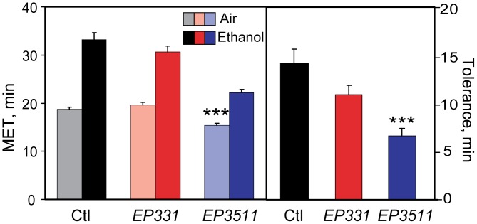
In mammals, both SAP97 and PSD95 have been shown to interact with the C-terminus of the NMDAR via their PDZ domains [28], [29]. Recent studies have shown that SAP97 plays an important role in cell surface trafficking and membrane clustering of NMDAR’s [32]. Numerous studies in rodents have demonstrated that compensatory synaptic changes occur in response to the inhibitory actions of ethanol on NMDAR complexes and that these effects may mediate synaptic plasticity involved in the development of ethanol tolerance [17]. In addition, it has been shown that NMDARs function in Drosophila olfactory learning and memory consolidation [42]. Hence, we hypothesized that a potential mechanism by which DlgS97 could regulate ethanol tolerance was through modulation of NMDAR function. To investigate this, we quantified ethanol tolerance in two hypomorphic mutants of Drosophila Nmdar1, EP3511 and EP331, caused by two independent P element insertion mutations in the Nmdar1 gene. These mutants have reduced levels of dNR1 protein, and display olfactory learning deficits [42]. Development of rapid tolerance to ethanol was significantly reduced, particularly in the EP3511 mutant (Fig. 5). Development of chronic tolerance was likewise reduced (Fig. S6). These finding are consistent with NMDARs in flies mediating synaptic and behavioral plasticity to ethanol exposure as has been reported in mammals [17].
Figure 5. A hypomorphic mutant of Nmdar1 exhibits reduced ethanol tolerance.
Ethanol sensitivity (left panel) and rapid tolerance (right panel) were quantified using the inebriometer in 2 different homozygous insertion mutants in Nmdar1, EP331 and EP3511, compared to control flies. The EP3511 mutant showed a highly significant reduction in tolerance, and also exhibited increased sensitivity (***, [F(2,20) = 21.338, p<0.001], one-way ANOVA with post hoc Holm-Sidak; n = 7 or 8). A second mutant in Nmdar1, EP331, showed a trend towards reduced tolerance (p = 0.057; n = 8). Dark colored bars represent inebriometer METs of flies pre-exposed to ethanol vapor, and light bars represent METs of parallel samples for each genotype pre-exposed to humidified air.
Molecular Complex Formation between DlgS97 and NMDAR1
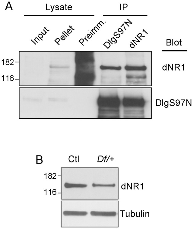
Ethanol-induced NMDAR-mediated synaptic plasticity involves synaptic remodeling that involves postsynaptic scaffolding molecules [17]. Moreover, mammalian homologs of DlgS97 interact with NMDARs and regulate subcellular targeting and synaptic plasticity mediated by NMDARs [28], [29], [54]. Therefore, we hypothesized that DlgS97 may form a complex with NMDAR1 and that its role in ethanol tolerance may be mediated through modification of NMDAR complexes in response to the acute effects of ethanol. Overall domain structures of both dNR1 and dNR2 proteins in Drosophila resemble those of their mammalian homologs. Sequence analysis of dNR1 revealed the presence of a type II PDZ domain binding motif, L(V)V, at the extreme C-terminus, which is absent from the dNR2 C-terminus. Hence, we focused on testing dNR1 interaction with DlgS97. We developed an anti-dNR1 antibody specific to the C-terminal domain of dNR1 (cytosolic region), that recognizes a single ∼130 kDa protein (Fig. 6B). We established the specificity of this antibody by using adult head extracts of flies heterozygous for a dNR1 deficiency, NR190B.0 in which the dNR1 gene is deleted [43]. The level of the putative dNR1 protein species was significantly reduced in dNR190B.0 heterozygotes (Fig. 6B, lane 2; homozygotes are unviable) in comparison to control flies (Fig. 6B, lane 1). We used this antibody to test whether DlgS97 formed a complex with dNR1 by immunoprecipitation of protein extracts of adult fly heads. As shown in Figure 7B, anti-dNR1 antibody, but not the cognate preimmune serum, co-precipitated DlgS97. In addition, anti-DlgS97N antibody co-precipitated dNR1, suggesting that dNR1 and DlgS97 form a complex in the adult fly brain as has been observed in the mammalian brain [31].
Figure 6. DlgS97 and NMDAR1 form a stable complex in vivo.
(A) Immunoprecipitation of a complex containing dNR1 and DlgS97 from adult head extracts. Proteins were immunoprecipitated with pre-immune control (lane 2), anti-DlgS97 antibody (lane 3) and anti-dNR1 antibody (lane 4). Input lane represents 5% of the total lysates. (B) Western blot analysis of the anti-dNR1 antibody using control (lane 1) and dNR1 heterozygous deficiency NR190B.0 flies (lane 2).
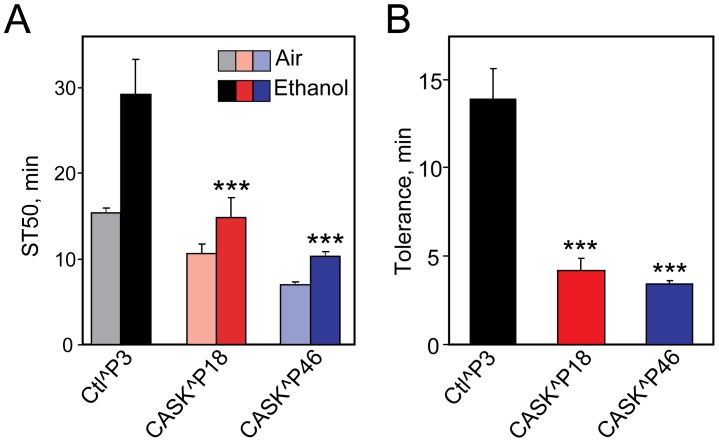
Figure 7. Hypomorphic mutants of Drosophila CASK display reduced ethanol tolerance.
Two different hypomorphic mutants of Drosophila CASK (CASKP18 and CASKP46) were tested for development of rapid tolerance, compared to an isogenic (CASK+) control. Time for 50% of each sample population of 25–30 flies to become sedated (ST50) was determined (panel A) and tolerance was quantified as the difference in ST50 between flies pre-exposed to ethanol and those pre-exposed to humidified air (panel B). There was a highly significant effect of genotype on both sensitivity and tolerance, as indicated (***, [F(3,22) = 25.241, p<0.001], one-way ANOVA with post hoc Holm-Sidak; n = 6–8).
Mutants in Camguk/Caki, Another Component of the Nmdar1 Scaffold, Exhibit Reduced Ethanol Tolerance
The Drosophila homolog of human CASK (also known as Caki or Camguk) is another L27 domain-containing molecular scaffold implicated in regulating synaptic plasticity [55]. The L27 domain of mammalian Camguk has been shown to interact with the L27 domain of SAP97 and this interaction is important for the subcellular localization of SAP97 [22]. Hence, we hypothesized that Camguk may play a role in the development of tolerance to ethanol. Mutants of Drosophila CASK were recently generated and characterized [44]. We tested these fly CASK hypomorphic mutants for ethanol tolerance, and found that they exhibited severely reduced tolerance, as well as increased sensitivity to ethanol sedation (Fig. 7). We suggest that the fly CASK homolog, along with DlgS97, is implicated in the adaptive molecular changes at synapses, likely involving NMDA receptor modulation, underlying the development of ethanol tolerance.
Mice with Reduced Levels of SAP97 Display Deficits in Rapid Tolerance
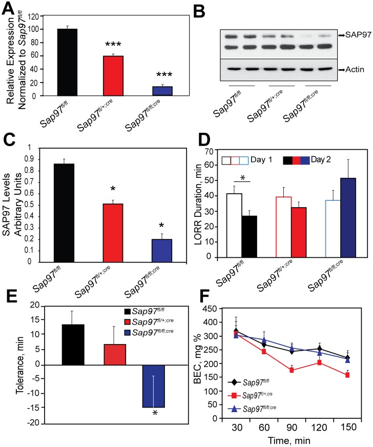
SAP97 is the mammalian homolog of Drosophila DlgS97 [24]. The loss of SAP97 function through development results in abnormalities in craniofacial development and perinatal lethality. Hence, we examined the development of rapid tolerance to ethanol in mice carrying a floxed Sap97 allele [47]. These mice were crossed with mice carrying the estrogen receptor (ESR)-Cre transgene. In this inducible-Cre transgenic mouse line, the Cre recombinase is fused to a mutant form of the mouse ESR ligand-binding domain that does not bind endogenous ligand at physiological concentrations, but binds the synthetic ligand Tamoxifen (TM). Upon injection of TM, the Cre-ESR fusion protein translocates to the nucleus and mediates deletion of the floxed allele resulting in temporal control of gene deletion [56]. To confirm that injection of TM did indeed delete the floxed allele, we examined Sap97 mRNA levels in the brains of Sap97fl/fl, Sap97fl/+;cre and Sap97fl/fl;cre mice 3–4 weeks after the final injection of TM. QPCR revealed that Sap97 mRNA levels were reduced by about 50% in Sap97fl/+;cre mice and by 90% in Sap97fl/fl;cre mice (Fig. 8A) in comparison to Sap97fl/fl mice. We also examined SAP97 protein levels in whole brain protein extracts prepared from Sap97fl/fl, Sap97fl/+;cre, and Sap97fl/fl;cre mice (Fig. 8B and C). Both heterozygous and homozygous mice carrying the Cre transgene showed significantly less SAP97 expression than Sap97fl/fl mice. However, SAP97 expression was not completely eliminated even 3–4 weeks after TM injection as evidenced by the presence of a faint band corresponding to SAP97 in the Sap97fl/fl;cre mice.
Figure 8. Mice with reduced SAP97 levels do not develop rapid tolerance to ethanol.
(A) Sap97 mRNA levels were compared between Sap97fl/fl, Sap97fl/+;cre and Sap97fl/fl;cre mice 3–4 weeks after TM injection. Male and female mice were used in this experiment. One Way ANOVA revealed a significant difference in Sap97 levels between the three genotypes [F[2], [15] = 127.326, p<0.001]. Bonferroni post hoc test revealed that Sap97 mRNA levels were significantly reduced to 60% in Sap97fl/+;cre mice (***, p<0.001) and to 10% in Sap97fl/fl;cre mice in comparison to Sap97fl/fl mice (***, p<0.001). n = 9 (Sap97fl/fl), 4 (Sap97fl/+;cre), and 5 (Sap97fl/fl;cre). (B) Representative western blot showing reduction in SAP97 protein levels in whole brain protein extracts of Sap97fl/fl, Sap97fl/+;cre and Sap97fl/fl;cre mice 3–4 weeks after TM injection. (C) Quantification of results in B. One-Way ANOVA revealed a significant main effect of genotype [F[2,9] = 55.931, p<0.001]. Bonferroni posthoc test revealed that Sap97fl/+;cre and Sap97fl/fl;cre mice expressed significantly less SAP97 in comparison to Sap97fl/fl mice (***, p<0.001). n = 4/genotype. D) Ethanol-induced LORR was measured in Sap97fl/fl, Sap97fl/+;cre and Sap97fl/fl;cre mice on two consecutive days. Two-Way repeated measures (RM) ANOVA detected a significant genotype×day interaction [F[2], [18] = 4.369, p = 0.029]. Bonferroni posthoc test revealed that the duration of LORR was significantly lower on Day 2 in comparison to Day 1 in Sap97fl/fl (*, p<0.05), but not Sap97fl/+;cre or Sap97fl/fl;cre mice, n = 13 (Sap97fl/fl ), 12 (Sap97fl/+;cre), and 10 (Sap97fl/fl;cre). (E) Tolerance scores, obtained by subtracting duration of LORR on Day 1 from Day 2, are shown. One–Way ANOVA revealed a significant effect of genotype on the development of tolerance [F(2,32) = 4.072, p<0.05]. Bonferroni post hoc test suggested that Sap97fl/fl mice developed significantly more tolerance to ethanol’s sedative hypnotic effects than Sap97fl/fl;cre mice, (*, p<0.05). (F) Ethanol metabolism curves are shown for the three genotypes. Two-Way RM ANOVA revealed a significant main effect of time [F [4,105] = 19.870, p<0.001] but no genotype [F [2,105] = 1.470, P>0.05] or time×genotype interaction [F [8,105] = 0.916, p>0.05] suggesting that all three genotypes metabolized ethanol at the same rate. n = 10 for (Sap97fl/fl, Sap97fl/fl;cre), n = 9 (Sap97fl/+;cre).
We compared the development of rapid tolerance to the sedative hypnotic effects of ethanol in these mice 3–4 weeks after TM injection. Mice were injected with 4 g/kg ethanol (i.p.) and the duration of the loss of righting (LORR) was recorded on Day 1. Twenty-four hours later, these mice were again injected with the same dose of ethanol and LORR was recorded as on Day 1. Significant differences were not observed in the duration of LORR between the genotypes on Day 1, suggesting that reduced SAP97 levels did not affect sensitivity to ethanol’s sedative/hypnotic effects. The duration of LORR in Sap97fl/fl mice was 15 minutes longer on Day 2 than on Day 1 (Fig. 8D and E), suggesting the development of robust rapid tolerance to the sedative/hypnotic effects of ethanol. However, we did not observe significant differences in the duration of LORR in Sap97fl/+;cre and Sap97fl/fl;cre mice between Day 1 and Day 2, suggesting that these mice do not develop rapid tolerance to ethanol. Figure 8E shows the LORR data as a tolerance score obtained by subtracting the duration of LORR on Day 1 from Day 2. Sap97fl/fl;cre mice developed significantly more tolerance to ethanol than Sap97fl/fl mice (Fig. 8E). We also examined metabolism of 4 g/kg ethanol (Fig. 8F). Statistically significant differences were not detected in the rate of metabolism of ethanol between the genotypes, suggesting that the observed differences in rapid tolerance were not due to altered metabolism of ethanol. In summary, adult mice with reduced expression of SAP97 showed defective development of rapid tolerance to the sedating effects of ethanol, arguing for a conserved role for SAP97 in ethanol tolerance.
Discussion
Here we describe a role for Drosophila DlgS97 and its murine homolog SAP97 in the development of tolerance to the sedating effects of ethanol. Our biochemical and behavioral data suggest NMDAR-mediated synaptic plasticity is a potential mechanism by which DlgS97 regulates ethanol tolerance. Furthermore, we find that mice in which the Sap97 gene has been deleted in adults show deficits in the development of rapid tolerance to the sedative/hypnotic effects of ethanol.
DlgS97 has a Conserved Role in Regulating Ethanol Tolerance
The Dlg-MAGUK family of scaffolding proteins, especially PSD-95 and SAP97, play critical roles in synaptic plasticity by participating in remodeling the molecular architecture at the synapse in an experience-and activity-dependent manner. As we observed, the ethanol tolerance defect of the intol fly mutant was only rescued upon transgenic expression of DlgS97, not DlgA, suggesting that the synaptic changes involved in developing ethanol tolerance are mediated by DlgS97.
The crucial structural difference between DlgA and DlgS97 is their N-terminus, where DlgS97 contains an L27 domain that is absent from DlgA. Biochemical and cell biological studies of the mammalian homologs of Dlg proteins, SAP97 and PSD-95, reported the importance of the L27 domain in targeting GluR1 and AMPA receptors by mediating oligomerization of SAP97 and the formation of a complex between SAP97 and PSD-95 [57], [58], [59].
The L27 domain also links SAP97 to its role in activity-dependent NMDAR and AMPAR signaling. RNAi knockdown of endogenous SAP97 reduces surface expression of both GluR1 and GluR2 and inhibits both AMPA and NMDA excitatory post synaptic currents (EPSCs) [57], demonstrating a broader role than PSD-95 in the maintenance of synaptic function. A recent study suggests that the L27 domain of SAP97 regulates synaptic plasticity by sequestering NMDARs at extrasynaptic sites [54]. The L27 domain of SAP97 is important for activity dependent regulation of AMPAR through activation of CamKII [60]. Furthermore, the L27 domain of DlgS97 interacts with Camguk and regulates CaMKII autophosphorylation, which has been shown to be a critical and evolutionarily conserved determinant of synaptic plasticity [55], [61], [62].
Consistent with the behavioral data obtained in flies, we found mice with targeted disruption of Sap97 fail to develop tolerance to the sedative/hypnotic effects of ethanol. We chose to measure the development of tolerance to ethanol’s sedative/hypnotic effects using the LORR assay [33], [34], [35] because of the similarity of this behavioral paradigm to the one used to measure ethanol tolerance in flies. We achieved acute knockdown of SAP97 levels by crossing mice harboring the floxed Sap97 allele with mice carrying the ESR-Cre transgene. Administering tamoxifen (TM) to these mice results in translocation of ESR-Cre into the nucleus and subsequent deletion of the floxed allele. Since TM is not administered until adulthood, we hypothesized that this approach would minimize the development of compensatory changes in the expression of other MAGUK family proteins in response to reduced SAP97 levels. Deletion of Sap97 does not affect the duration of LORR on day 1 (ethanol-naïve mice), suggesting that SAP97 does not affect initial hypnotic sensitivity to ethanol. This finding differs from the observation that intol flies showed a slightly lower sensitivity to the sedating effects of ethanol (it should be noted that the intol mutation is not conditional; we were unable to generate an adult-specific fly intol mutant). Sap97fl/fl mice developed robust tolerance to the sedative/hypnotic effects of ethanol as evidenced by decreased duration of LORR on day 2 compared to day 1. In contrast, Sap97fl/fl ;cre mice failed to develop rapid tolerance to ethanol’s sedative/hypnotic effects. Sap97fl/+;cre mice displayed a trend towards reduced tolerance, but these results did not reach statistical significance. In summary, these results implicate a novel and conserved role for SAP97 in mediating the development of ethanol tolerance.
Brain Regions Implicated in Ethanol Tolerance
Previous studies showed that the ability to develop tolerance was significantly reduced in Drosophila mutants with structural and functional abnormalities in the central complex and mushroom body [4]. In the current study, we noted that behavioral rescue of the intol mutant was associated with strong expression of UAS-DlgS97 in the mushroom body. This in turn suggests that DlgS97 protein expression in the mushroom body is relevant for normal development of ethanol tolerance. Consistent with this notion, endogenous DlgS97 is highly expressed in the mushroom body. A previous study from our laboratory examined fly learning and memory “enhancer trap” mutants for their ability to develop ethanol tolerance; indeed several of the mutants with expression in the mushroom body had defective tolerance [7]. The fact that the behavioral rescue we observed for intol; GAL4c747;UAS-DlgS97 was only partial is consistent with other brain regions also being involved, which is supported by previous studies [63].
Molecular Interactions of DlgS97 and Ethanol Tolerance
The results of the present study indicate that in the fly brain, DlgS97 and NMDARs form a functional complex. The formation of a molecular complex between DlgS97 and NMDARs may play an important role in the clustering and subcellular targeting of NMDARs at the synapse and consequently impact synaptic plasticity and the development of tolerance to ethanol. In mammals, SAP97 is known to be involved mainly in targeting and clustering AMPARs via interaction through PDZ domains [59], [64]. However, SAP97 also binds to the NR2A and NR2B subunits of NMDA receptors [28], [29], [65]. The L27 domain of DlgS97 also interacts with Camguk, the Drosophila CASK homolog. Camguk has been shown to bind to the regulatory subunit of CaMKII in an ATP-dependent manner. Camguk modulates synaptic plasticity by regulating CaMKII autophosphorylation in an activity-dependent manner [55], [61]. The interactions of DlgS97 with Camguk and dNR1 is likely important for ethanol tolerance, given that fly Camguk and dNR1 mutants, like DlgS97 mutants, showed defective tolerance development. Our results suggest that a molecular scaffold comprised of DlgS97, Camguk, and dNR1 plays an important role in the development of tolerance to ethanol.
In summary, we have uncovered a novel role for DlgS97 in the development of tolerance to ethanol’s sedative/hypnotic effects. Strikingly, we found that mice with reduced expression of SAP97 display deficits in the development of rapid tolerance to ethanol. This further supports the notion that the molecular machinery that underlies development of tolerance to ethanol is largely conserved between flies and mammals, and identifies particular molecular players and interactions for future investigation.
Supporting Information
The intolerant mutant flies exhibit normal ethanol absorption and metabolism. Control (2202U) and intol mutant flies were grown and collected as for behavioral experiments, pre-exposed to ethanol vapor or humidified air, allowed to recover and re-exposed all to ethanol vapor, and snap-frozen at time indicated by arrow in schematic. Frozen flies were processed and ethanol content quantified. No significant effect of either genotype or pre-exposure condition (air vs. ethanol vapor) was seen on ethanol content of flies, nor was there a significant interaction between genotype and pre-exposure condition (two-way ANOVA; n = 4).
(TIF)
A longer ethanol pre-exposure does not correct the intol mutant tolerance deficit. Parental (2202U) and intol mutant flies were pre-exposed to ethanol vapor or to humidified air (control) for 30, 40 or 50 min as indicated and allowed to recover for ∼3.5 hr. All samples were then exposed to ethanol vapor (110∶40 relative flow ethanol vapor:humidified air), and the number of flies unable to stand at 2-min intervals during exposure was counted. Time for 50% of each sample population of 25–30 flies to become sedated (ST50) was determined (panel A) and tolerance was quantified as the difference in ST50 between flies pre-exposed to ethanol and those pre-exposed to humidified air (panel B). There was a highly significant effect of genotype on tolerance, as indicated (***, p<0.001), and also of pre-exposure duration, as well as a significant interaction between genotype and pre-exposure duration (two-way ANOVA; n = 4 (30 min and 50 min pre-exposure) or 8 (40 min pre-exposure).
(TIF)
The intol mutation is recessive. Female flies (the intol mutation is X-linked) which were either intol+/intol+ (Ctl), homozygous mutant (intol/intol) or heterozygous (intol/+) were pre-exposed to ethanol vapor (30 min at 60/40 relative flow rate; darker bars, left panel) or humidified air (lighter bars, left panel), allowed to recover for 3.5 hr, and assayed in the inebriometer. Tolerance was quantified as the difference in MET between flies pre-exposed to ethanol vapor vs. humidified air (right panel). A tolerance defect was seen for intol/intol homozygous flies, while intol/+ flies were indistinguishable from Ctl (***, p<0.001; n = 4 (Ctl), 6 (intol/intol) or 8 (intol/+)). No significant effect of genotype on initial sensitivity (MET of air pre-exposed flies) was detected in this experiment (p>0.3).
(TIF)
Mutations in dlg1 cause reduced chronic tolerance. The intol mutant, and 2 other independently identified dlg1 mutants, exhibit reduced chronic tolerance development compared to the genetic background strain, 2202U (“Ctl”) (*, p<0.05; ***, p<0.001; one-way ANOVA with post hoc Holm-Sidak test; n = 7 (Ctl), 6 (NP0768, NP1102, EY05003), 11 (intol) or 4 (NP4134)). Chronic tolerance was measured in flies which were pre-exposed overnight to a low, non-sedating concentration of ethanol vapor (10∶80 relative units ethanol vapor: humidified air) or to humidified air alone, essentially as previously described [6].
(TIF)
Dlg1 mutant larvae show loss of DlgS97 while DlgA expression is intact. (A) Protein expression detected by immunofluorescence at wild-type and mutant 3rd instar larval NMJ. Muscle 4 at abdominal segment A3 of control and dlg mutant larvae was triple-labeled with anti-dSAP97 (DlgS97N), anti-pan-Dlg antibody (DlgPDZ) and Cy3-HRP (HRP). Each image represents a stack of 15 optical sections taken at 0.5 µm steps. (B) Western blot analysis of body wall muscles from the wild type control and the dlg1 mutants 15779, intol, NP0768, NP1102, and NP4314. Immunoblot analysis of protein from 3rd instar larvae was performed using anti-pan-Dlg antibody (DlgPDZ) and DlgS97-specific antibody (DlgS97N).
(TIF)
Hypomorphic mutants of Nmdar1 exhibit reduced chronic ethanol tolerance. Ethanol sensitivity (A) and chronic tolerance (B) were quantified using the inebriometer for 2 different homozygous insertion mutants in Nmdar1, EP331 and EP3511, compared to isogenic background control flies. The EP3511 mutant showed a highly significant reduction in tolerance, and also exhibited increased sensitivity (p<0.001, one-way ANOVA with post hoc Holm-Sidak; n = 9 or 10). A second mutant in Nmdar1, EP331, also showed significantly reduced tolerance (p<0.001), but no alteration in sensitivity (n = 9). In panel A, dark colored bars represent inebriometer METs of flies pre-exposed to ethanol vapor, and light bars represent METs of parallel samples for each genotype pre-exposed to humidified air.
(TIF)
(DOC)
Acknowledgments
The authors are grateful to Francois Schweisguth and Yahanns Bellaiche for early collaborations in elucidating the cell biological and functional analysis of DlgS97 at the larval neuromuscular junctions, Graeme Davis for performing the electrophysiological analysis of dlg mutant larvae, Vivian Budnik for providing the UAS-Dlg-GFP transgene, Uli Thomas for providing the UAS-DlgS97-GFP construct. We thank members of the Heberlein lab for valuable discussions.
Funding Statement
This work was supported by the State of California (63-55) and National Institutes of health/National Institute on Alcohol Abuse and Alcoholism grants to UH and Ruth Kirschstein National Research Service Award (F32AA15470-02) to SL. The funders had no role in study design, data collection and analysis, decision to publish, or preparation of the manuscript.
References
- 1. Chandler LJ, Harris RA, Crews FT (1998) Ethanol tolerance and synaptic plasticity. Trends Pharmacol Sci 19: 491–495. [DOI] [PubMed] [Google Scholar]
- 2. Fadda F, Rossetti ZL (1998) Chronic ethanol consumption: from neuroadaptation to neurodegeneration. Prog Neurobiol 56: 385–431. [DOI] [PubMed] [Google Scholar]
- 3. Devineni AV, McClure K, Guarnieri D, Corl A, Wolf F, et al. The genetic relationships between ethanol preference, acute ethanol sensitivity, and ethanol tolerance in Drosophila melanogaster. Fly (Austin) 5: 191–199. [DOI] [PMC free article] [PubMed] [Google Scholar]
- 4. Scholz H, Ramond J, Singh CM, Heberlein U (2000) Functional ethanol tolerance in Drosophila. Neuron 28: 261–271. [DOI] [PubMed] [Google Scholar]
- 5. Scholz H, Franz M, Heberlein U (2005) The hangover gene defines a stress pathway required for ethanol tolerance development. Nature 436: 845–847. [DOI] [PMC free article] [PubMed] [Google Scholar]
- 6. Berger KH, Heberlein U, Moore MS (2004) Rapid and chronic: two distinct forms of ethanol tolerance in Drosophila. Alcohol Clin Exp Res 28: 1469–1480. [DOI] [PubMed] [Google Scholar]
- 7. Berger KH, Kong EC, Dubnau J, Tully T, Moore MS, et al. (2008) Ethanol sensitivity and tolerance in long-term memory mutants of Drosophila melanogaster. Alcohol Clin Exp Res 32: 895–908. [DOI] [PMC free article] [PubMed] [Google Scholar]
- 8.Krishnan HR, Al-Hasan YM, Pohl JB, Ghezzi A, Atkinson NS A Role for Dynamin in Triggering Ethanol Tolerance. Alcohol Clin Exp Res. [DOI] [PMC free article] [PubMed]
- 9. Kong EC, Allouche L, Chapot PA, Vranizan K, Moore MS, et al. Ethanol-regulated genes that contribute to ethanol sensitivity and rapid tolerance in Drosophila. Alcohol Clin Exp Res 34: 302–316. [DOI] [PMC free article] [PubMed] [Google Scholar]
- 10. Dimitrijevic N, Dzitoyeva S, Satta R, Imbesi M, Yildiz S, et al. (2005) Drosophila GABA(B) receptors are involved in behavioral effects of gamma-hydroxybutyric acid (GHB). Eur J Pharmacol 519: 246–252. [DOI] [PubMed] [Google Scholar]
- 11. Cowmeadow RB, Krishnan HR, Atkinson NS (2005) The slowpoke gene is necessary for rapid ethanol tolerance in Drosophila. Alcohol Clin Exp Res 29: 1777–1786. [DOI] [PubMed] [Google Scholar]
- 12. Cowmeadow RB, Krishnan HR, Ghezzi A, Al'Hasan YM, Wang YZ, et al. (2006) Ethanol tolerance caused by slowpoke induction in Drosophila. Alcohol Clin Exp Res 30: 745–753. [DOI] [PubMed] [Google Scholar]
- 13. Bhandari P, Kendler KS, Bettinger JC, Davies AG, Grotewiel M (2009) An assay for evoked locomotor behavior in Drosophila reveals a role for integrins in ethanol sensitivity and rapid ethanol tolerance. Alcohol Clin Exp Res 33: 1794–1805. [DOI] [PMC free article] [PubMed] [Google Scholar]
- 14. Urizar NL, Yang Z, Edenberg HJ, Davis RL (2007) Drosophila homer is required in a small set of neurons including the ellipsoid body for normal ethanol sensitivity and tolerance. J Neurosci 27: 4541–4551. [DOI] [PMC free article] [PubMed] [Google Scholar]
- 15. Li C, Zhao X, Cao X, Chu D, Chen J, et al. (2008) The Drosophila homolog of jwa is required for ethanol tolerance. Alcohol Alcohol 43: 529–536. [DOI] [PubMed] [Google Scholar]
- 16. Gardoni F (2008) MAGUK proteins: new targets for pharmacological intervention in the glutamatergic synapse. Eur J Pharmacol 585: 147–152. [DOI] [PubMed] [Google Scholar]
- 17. Nagy J (2008) Alcohol related changes in regulation of NMDA receptor functions. Curr Neuropharmacol 6: 39–54. [DOI] [PMC free article] [PubMed] [Google Scholar]
- 18. Alvestad RM, Grosshans DR, Coultrap SJ, Nakazawa T, Yamamoto T, et al. (2003) Tyrosine dephosphorylation and ethanol inhibition of N-Methyl-D-aspartate receptor function. J Biol Chem 278: 11020–11025. [DOI] [PubMed] [Google Scholar]
- 19. Anders DL, Blevins T, Sutton G, Chandler LJ, Woodward JJ (1999) Effects of c-Src tyrosine kinase on ethanol sensitivity of recombinant NMDA receptors expressed in HEK 293 cells. Alcohol Clin Exp Res 23: 357–362. [PubMed] [Google Scholar]
- 20. Maldve RE, Zhang TA, Ferrani-Kile K, Schreiber SS, Lippmann MJ, et al. (2002) DARPP-32 and regulation of the ethanol sensitivity of NMDA receptors in the nucleus accumbens. Nat Neurosci 5: 641–648. [DOI] [PubMed] [Google Scholar]
- 21. Krystal JH, Petrakis IL, Mason G, Trevisan L, D'Souza DC (2003) N-methyl-D-aspartate glutamate receptors and alcoholism: reward, dependence, treatment, and vulnerability. Pharmacol Ther 99: 79–94. [DOI] [PubMed] [Google Scholar]
- 22. Lee S, Fan S, Makarova O, Straight S, Margolis B (2002) A novel and conserved protein-protein interaction domain of mammalian Lin-2/CASK binds and recruits SAP97 to the lateral surface of epithelia. Mol Cell Biol 22: 1778–1791. [DOI] [PMC free article] [PubMed] [Google Scholar]
- 23. Mendoza C, Olguin P, Lafferte G, Thomas U, Ebitsch S, et al. (2003) Novel isoforms of Dlg are fundamental for neuronal development in Drosophila. J Neurosci 23: 2093–2101. [DOI] [PMC free article] [PubMed] [Google Scholar]
- 24. Sheng M, Sala C (2001) PDZ domains and the organization of supramolecular complexes. Annu Rev Neurosci 24: 1–29. [DOI] [PubMed] [Google Scholar]
- 25. Thomas U, Kobler O, Gundelfinger ED The Drosophila larval neuromuscular junction as a model for scaffold complexes at glutamatergic synapses: benefits and limitations. J Neurogenet 24: 109–119. [DOI] [PubMed] [Google Scholar]
- 26. Mendoza-Topaz C, Urra F, Barria R, Albornoz V, Ugalde D, et al. (2008) DLGS97/SAP97 is developmentally upregulated and is required for complex adult behaviors and synapse morphology and function. J Neurosci 28: 304–314. [DOI] [PMC free article] [PubMed] [Google Scholar]
- 27. Fujita A, Kurachi Y (2000) SAP family proteins. Biochem Biophys Res Commun 269: 1–6. [DOI] [PubMed] [Google Scholar]
- 28. Bassand P, Bernard A, Rafiki A, Gayet D, Khrestchatisky M (1999) Differential interaction of the tSXV motifs of the NR1 and NR2A NMDA receptor subunits with PSD-95 and SAP97. Eur J Neurosci 11: 2031–2043. [DOI] [PubMed] [Google Scholar]
- 29. Niethammer M, Kim E, Sheng M (1996) Interaction between the C terminus of NMDA receptor subunits and multiple members of the PSD-95 family of membrane-associated guanylate kinases. J Neurosci 16: 2157–2163. [DOI] [PMC free article] [PubMed] [Google Scholar]
- 30. Cai C, Coleman SK, Niemi K, Keinanen K (2002) Selective binding of synapse-associated protein 97 to GluR-A alpha-amino-5-hydroxy-3-methyl-4-isoxazole propionate receptor subunit is determined by a novel sequence motif. J Biol Chem 277: 31484–31490. [DOI] [PubMed] [Google Scholar]
- 31. Gardoni F, Mauceri D, Fiorentini C, Bellone C, Missale C, et al. (2003) CaMKII-dependent phosphorylation regulates SAP97/NR2A interaction. J Biol Chem 278: 44745–44752. [DOI] [PubMed] [Google Scholar]
- 32. Jeyifous O, Waites CL, Specht CG, Fujisawa S, Schubert M, et al. (2009) SAP97 and CASK mediate sorting of NMDA receptors through a previously unknown secretory pathway. Nat Neurosci 12: 1011–1019. [DOI] [PMC free article] [PubMed] [Google Scholar]
- 33. Yang X, Oswald L, Wand G (2003) The cyclic AMP/protein kinase A signal transduction pathway modulates tolerance to sedative and hypothermic effects of ethanol. Alcohol Clin Exp Res 27: 1220–1225. [DOI] [PubMed] [Google Scholar]
- 34.Sato Y, Seo N, Kobayashi E (2006) Ethanol-induced hypnotic tolerance is absent in N-methyl-D-aspartate receptor epsilon 1 subunit knockout mice. Anesth Analg 103: 117–120, table of contents. [DOI] [PubMed]
- 35. Wu PH, Liu JF, Wu WL, Lanca AJ, Kalant H (1996) Development of alcohol tolerance in the rat after a single exposure to combined treatment with arginine8-vasopressin and ethanol. J Pharmacol Exp Ther 276: 1283–1291. [PubMed] [Google Scholar]
- 36. Brand AH, Perrimon N (1993) Targeted gene expression as a means of altering cell fates and generating dominant phenotypes. Development 118: 401–415. [DOI] [PubMed] [Google Scholar]
- 37. Corl AB, Berger KH, Ophir-Shohat G, Gesch J, Simms JA, et al. (2009) Happyhour, a Ste20 family kinase, implicates EGFR signaling in ethanol-induced behaviors. Cell 137: 949–960. [DOI] [PubMed] [Google Scholar]
- 38. Dubnau J, Chiang AS, Grady L, Barditch J, Gossweiler S, et al. (2003) The staufen/pumilio pathway is involved in Drosophila long-term memory. Curr Biol 13: 286–296. [DOI] [PubMed] [Google Scholar]
- 39. Woods DF, Bryant PJ (1989) Molecular cloning of the lethal(1)discs large-1 oncogene of Drosophila. Dev Biol 134: 222–235. [DOI] [PubMed] [Google Scholar]
- 40. Koh YH, Popova E, Thomas U, Griffith LC, Budnik V (1999) Regulation of DLG localization at synapses by CaMKII-dependent phosphorylation. Cell 98: 353–363. [DOI] [PMC free article] [PubMed] [Google Scholar]
- 41. Bachmann A, Timmer M, Sierralta J, Pietrini G, Gundelfinger ED, et al. (2004) Cell type-specific recruitment of Drosophila Lin-7 to distinct MAGUK-based protein complexes defines novel roles for Sdt and Dlg-S97. J Cell Sci 117: 1899–1909. [DOI] [PubMed] [Google Scholar]
- 42. Xia S, Miyashita T, Fu TF, Lin WY, Wu CL, et al. (2005) NMDA receptors mediate olfactory learning and memory in Drosophila. Curr Biol 15: 603–615. [DOI] [PMC free article] [PubMed] [Google Scholar]
- 43. Venkatesh K, Hasan G (1997) Disruption of the IP3 receptor gene of Drosophila affects larval metamorphosis and ecdysone release. Curr Biol 7: 500–509. [DOI] [PubMed] [Google Scholar]
- 44. Slawson JB, Kuklin EA, Ejima A, Mukherjee K, Ostrovsky L, et al. Central regulation of locomotor behavior of Drosophila melanogaster depends on a CASK isoform containing CaMK-like and L27 domains. Genetics 187: 171–184. [DOI] [PMC free article] [PubMed] [Google Scholar]
- 45. Moore MS, DeZazzo J, Luk AY, Tully T, Singh CM, et al. (1998) Ethanol intoxication in Drosophila: Genetic and pharmacological evidence for regulation by the cAMP signaling pathway. Cell 93: 997–1007. [DOI] [PubMed] [Google Scholar]
- 46. Cohan FM, Hoffmann AA (1986) Genetic divergence under uniform selection. II. Different responses to selection for knockdown resistance to ethanol among Drosophila melanogaster populations and their replicate lines. Genetics 114: 145–164. [DOI] [PMC free article] [PubMed] [Google Scholar]
- 47. Zhou W, Zhang L, Guoxiang X, Mojsilovic-Petrovic J, Takamaya K, et al. (2008) GluR1 controls dendrite growth through its binding partner, SAP97. J Neurosci 28: 10220–10233. [DOI] [PMC free article] [PubMed] [Google Scholar]
- 48. Carnicella S, Amamoto R, Ron D (2009) Excessive alcohol consumption is blocked by glial cell line-derived neurotrophic factor. Alcohol 43: 35–43. [DOI] [PMC free article] [PubMed] [Google Scholar]
- 49. Wolf FW, Rodan AR, Tsai LT, Heberlein U (2002) High-resolution analysis of ethanol-induced locomotor stimulation in Drosophila. J Neurosci 22: 11035–11044. [DOI] [PMC free article] [PubMed] [Google Scholar]
- 50. Singh CM, Heberlein U (2000) Genetic control of acute ethanol-induced behaviors in Drosophila. Alcohol Clin Exp Res 24: 1127–1136. [PubMed] [Google Scholar]
- 51. Doerks T, Bork P, Kamberov E, Makarova O, Muecke S, et al. (2000) L27, a novel heterodimerization domain in receptor targeting proteins Lin-2 and Lin-7. Trends Biochem Sci 25: 317–318. [DOI] [PubMed] [Google Scholar]
- 52. McGuire SE, Le PT, Davis RL (2001) The role of Drosophila mushroom body signaling in olfactory memory. Science 293: 1330–1333. [DOI] [PubMed] [Google Scholar]
- 53. Bachmann A, Kobler O, Kittel RJ, Wichmann C, Sierralta J, et al. A perisynaptic menage a trois between Dlg, DLin-7, and Metro controls proper organization of Drosophila synaptic junctions. J Neurosci 30: 5811–5824. [DOI] [PMC free article] [PubMed] [Google Scholar]
- 54. Li D, Specht CG, Waites CL, Butler-Munro C, Leal-Ortiz S, et al. SAP97 directs NMDA receptor spine targeting and synaptic plasticity. J Physiol 589: 4491–4510. [DOI] [PMC free article] [PubMed] [Google Scholar]
- 55. Hodge JJ, Mullasseril P, Griffith LC (2006) Activity-dependent gating of CaMKII autonomous activity by Drosophila CASK. Neuron 51: 327–337. [DOI] [PubMed] [Google Scholar]
- 56. Hayashi S, McMahon AP (2002) Efficient recombination in diverse tissues by a tamoxifen-inducible form of Cre: a tool for temporally regulated gene activation/inactivation in the mouse. Dev Biol 244: 305–318. [DOI] [PubMed] [Google Scholar]
- 57. Nakagawa T, Futai K, Lashuel HA, Lo I, Okamoto K, et al. (2004) Quaternary structure, protein dynamics, and synaptic function of SAP97 controlled by L27 domain interactions. Neuron 44: 453–467. [DOI] [PubMed] [Google Scholar]
- 58. Feng W, Long JF, Fan JS, Suetake T, Zhang M (2004) The tetrameric L27 domain complex as an organization platform for supramolecular assemblies. Nat Struct Mol Biol 11: 475–480. [DOI] [PubMed] [Google Scholar]
- 59. Cai C, Li H, Rivera C, Keinanen K (2006) Interaction between SAP97 and PSD-95, two Maguk proteins involved in synaptic trafficking of AMPA receptors. J Biol Chem 281: 4267–4273. [DOI] [PubMed] [Google Scholar]
- 60. Schluter OM, Xu W, Malenka RC (2006) Alternative N-terminal domains of PSD-95 and SAP97 govern activity-dependent regulation of synaptic AMPA receptor function. Neuron 51: 99–111. [DOI] [PubMed] [Google Scholar]
- 61. Lu CS, Hodge JJ, Mehren J, Sun XX, Griffith LC (2003) Regulation of the Ca2+/CaM-responsive pool of CaMKII by scaffold-dependent autophosphorylation. Neuron 40: 1185–1197. [DOI] [PubMed] [Google Scholar]
- 62. Lisman J, Schulman H, Cline H (2002) The molecular basis of CaMKII function in synaptic and behavioural memory. Nat Rev Neurosci 3: 175–190. [DOI] [PubMed] [Google Scholar]
- 63. Scholz H (2005) Influence of the biogenic amine tyramine on ethanol-induced behaviors in Drosophila. J Neurobiol 63: 199–214. [DOI] [PubMed] [Google Scholar]
- 64. Leonard AS, Davare MA, Horne MC, Garner CC, Hell JW (1998) SAP97 is associated with the alpha-amino-3-hydroxy-5-methylisoxazole-4-propionic acid receptor GluR1 subunit. J Biol Chem 273: 19518–19524. [DOI] [PubMed] [Google Scholar]
- 65. Wang L, Piserchio A, Mierke DF (2005) Structural characterization of the intermolecular interactions of synapse-associated protein-97 with the NR2B subunit of N-methyl-D-aspartate receptors. J Biol Chem 280: 26992–26996. [DOI] [PubMed] [Google Scholar]
Associated Data
This section collects any data citations, data availability statements, or supplementary materials included in this article.
Supplementary Materials
The intolerant mutant flies exhibit normal ethanol absorption and metabolism. Control (2202U) and intol mutant flies were grown and collected as for behavioral experiments, pre-exposed to ethanol vapor or humidified air, allowed to recover and re-exposed all to ethanol vapor, and snap-frozen at time indicated by arrow in schematic. Frozen flies were processed and ethanol content quantified. No significant effect of either genotype or pre-exposure condition (air vs. ethanol vapor) was seen on ethanol content of flies, nor was there a significant interaction between genotype and pre-exposure condition (two-way ANOVA; n = 4).
(TIF)
A longer ethanol pre-exposure does not correct the intol mutant tolerance deficit. Parental (2202U) and intol mutant flies were pre-exposed to ethanol vapor or to humidified air (control) for 30, 40 or 50 min as indicated and allowed to recover for ∼3.5 hr. All samples were then exposed to ethanol vapor (110∶40 relative flow ethanol vapor:humidified air), and the number of flies unable to stand at 2-min intervals during exposure was counted. Time for 50% of each sample population of 25–30 flies to become sedated (ST50) was determined (panel A) and tolerance was quantified as the difference in ST50 between flies pre-exposed to ethanol and those pre-exposed to humidified air (panel B). There was a highly significant effect of genotype on tolerance, as indicated (***, p<0.001), and also of pre-exposure duration, as well as a significant interaction between genotype and pre-exposure duration (two-way ANOVA; n = 4 (30 min and 50 min pre-exposure) or 8 (40 min pre-exposure).
(TIF)
The intol mutation is recessive. Female flies (the intol mutation is X-linked) which were either intol+/intol+ (Ctl), homozygous mutant (intol/intol) or heterozygous (intol/+) were pre-exposed to ethanol vapor (30 min at 60/40 relative flow rate; darker bars, left panel) or humidified air (lighter bars, left panel), allowed to recover for 3.5 hr, and assayed in the inebriometer. Tolerance was quantified as the difference in MET between flies pre-exposed to ethanol vapor vs. humidified air (right panel). A tolerance defect was seen for intol/intol homozygous flies, while intol/+ flies were indistinguishable from Ctl (***, p<0.001; n = 4 (Ctl), 6 (intol/intol) or 8 (intol/+)). No significant effect of genotype on initial sensitivity (MET of air pre-exposed flies) was detected in this experiment (p>0.3).
(TIF)
Mutations in dlg1 cause reduced chronic tolerance. The intol mutant, and 2 other independently identified dlg1 mutants, exhibit reduced chronic tolerance development compared to the genetic background strain, 2202U (“Ctl”) (*, p<0.05; ***, p<0.001; one-way ANOVA with post hoc Holm-Sidak test; n = 7 (Ctl), 6 (NP0768, NP1102, EY05003), 11 (intol) or 4 (NP4134)). Chronic tolerance was measured in flies which were pre-exposed overnight to a low, non-sedating concentration of ethanol vapor (10∶80 relative units ethanol vapor: humidified air) or to humidified air alone, essentially as previously described [6].
(TIF)
Dlg1 mutant larvae show loss of DlgS97 while DlgA expression is intact. (A) Protein expression detected by immunofluorescence at wild-type and mutant 3rd instar larval NMJ. Muscle 4 at abdominal segment A3 of control and dlg mutant larvae was triple-labeled with anti-dSAP97 (DlgS97N), anti-pan-Dlg antibody (DlgPDZ) and Cy3-HRP (HRP). Each image represents a stack of 15 optical sections taken at 0.5 µm steps. (B) Western blot analysis of body wall muscles from the wild type control and the dlg1 mutants 15779, intol, NP0768, NP1102, and NP4314. Immunoblot analysis of protein from 3rd instar larvae was performed using anti-pan-Dlg antibody (DlgPDZ) and DlgS97-specific antibody (DlgS97N).
(TIF)
Hypomorphic mutants of Nmdar1 exhibit reduced chronic ethanol tolerance. Ethanol sensitivity (A) and chronic tolerance (B) were quantified using the inebriometer for 2 different homozygous insertion mutants in Nmdar1, EP331 and EP3511, compared to isogenic background control flies. The EP3511 mutant showed a highly significant reduction in tolerance, and also exhibited increased sensitivity (p<0.001, one-way ANOVA with post hoc Holm-Sidak; n = 9 or 10). A second mutant in Nmdar1, EP331, also showed significantly reduced tolerance (p<0.001), but no alteration in sensitivity (n = 9). In panel A, dark colored bars represent inebriometer METs of flies pre-exposed to ethanol vapor, and light bars represent METs of parallel samples for each genotype pre-exposed to humidified air.
(TIF)
(DOC)


