Figure 1.
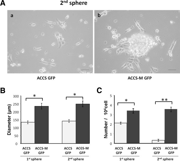
Cells with EMT alterations show sphere-forming ability. ACCS-GFP and ACCS-M GFP cells were cultured at a density of 5 × 10⁴ cells/mL in serum-free medium containing 40 ng/mL bFGF and 20 ng/mL EGF for floating culture for 10 days (primary spheres). For secondary spheres, primary spheres (day 10) were dissociated into single cells and further cultured at a density of 1 × 10⁴ cells/mL for 10 days. Spheres were observed under a phase contrast microscope (A). Sphere diameters were measured (B), and spheres with a diameter >100 μm were counted. Sphere numbers were standardized as sphere number/104 cells originally cultured (C) in each sphere period. Experiments were performed in triplicate, and the values were averaged. Bars indicate the standard deviation. Data significance was analyzed by Student’s t-test. *P < 0.05, **P < 0.01.

