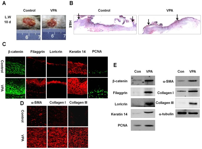
Figure 4. Effects of VPA on cutaneous wound healing in large wounds.
A full-thickness skin excision (diameter = 1.5 cm) was made on the backs of 8-week-old C3H mice, and 500 mM VPA was topically applied to the wounds daily. Tissues were excised from the wounded area and fixed in paraformaldehyde for immunohistochemistry or frozen in liquid nitrogen for Western blotting. (A) Representative gross images of wounded skin after 10-d VPA treatment. (B) Representative H&E stained tissues of wounded skin treated with or without VPA. Arrows represent the wound edge (original magnification ×40). (C) Immunohistochemical analysis of β-catenin, filaggrin, loricrin, keratin 14, or PCNA in the neo-epidermis of control and VPA-treated wounds (original magnification ×635). (D) Immunohistochemical analysis of α-SMA, collagen I, or collagen III in the control and VPA-treated wounds (original magnification ×635). (E) Western blot analysis of β-catenin, filaggrin, loricrin, keratin 14, PCNA, α-SMA, collagen I, collagen III, or α-tubulin in the control and VPA-treated wounds.

